Showing posts with label PET. Show all posts

March 1, 2014

Sarcoidosis on PET/CT


(A) MIP image from a PET/CT shows areas of FDG avidity in multiple lymph node stations including hilar, mediastinal, axillary, upper abdominal and groin regions. Note intense uptake of the spleen. 

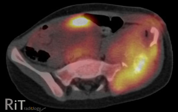
(B&C) Axial fused PET/CT images show intense FDG uptake within thoracic, axillary nodes and spleen. 

PET/CT: Three patterns of sarcoidosis
  1. Typical: Bilateral hilar uptake extending to the mediastinum with bilateral lung uptakes (PET and CT concordant lesions). This is found in the majority of cases (about 2/3)
  2. Discrepant: Multiple foci of uptake in and outside chest, along with splenic uptake (PET and CT discordant lesions). Fewer lesions are seen on CT than on PET. This pattern is the 2nd most common and is indistinguishable from malignancy (esp. metastasis, lymphoma)
  3. Multiple small FDG avid lung lesions: This pattern is similar to lung metastasis. Fortunately, it is the least common pattern. 
Our case: Biopsy-confirmed sarcoidosis involving the hilar, mediastinal, upper abdominal lymph nodes, and spleen. This follows the "discrepant" pattern (basically meaning that malignancy cannot be reliably distinguished)

References:
Alavi A, et al. Positron emission tomography imaging in nonmalignant thoracic disorders. Semin Nucl Med 2002;32:293-321.


January 11, 2013

Extraosseous Myeloma


Axial PET/CT images show a large homogeneous soft tissue mass in the lett buttock involving the gluteal muscles, which demonstrates significant FDG avidity. 


Extraosseous Myeloma

  • 10-16% of patients with multiple myeloma
  • Rising incidence possibly due to better imaging techniques and increased survival of patients with myeloma
  • More common in younger myeloma patients and more aggressive subtypes
  • Shorter survival and shorter progression-free survival (than patients without extraosseous involvement)
  • Has been integrated into an updated version of Durie-Salmon staging system

How to Image Patients with Myeloma

  • Skeletal survey (current minimum standard)
  • Whole-body PET/CT vs. whole-body MRI for newly diagnosed myeloma and with no findings or limited findings on skeletal survey (two different methods have been suggested by different authorities) to assess for occult involvement
Imaging Appearances
  • Soft tissue: nodules in subcutaneous tissues and muscles (like in our case)
  • Reticuloendothelial system: lymph node enlargement in multiple stations
  • Liver: hepatomegaly, low-attenuation liver lesions without enhancement
  • CNS: leptomengineal process 
  • Lungs: lung nodules, masses, interstitial infiltration
  • Kidneys: masses, diffuse enlargement
  • Peritoneum: masses
Reference:
Hall MN, Jagannathan JP, Ramaiya NH, et al. Imaging of extraosseous myeloma: CT, PET/CT and MRI features. AJR 2010; 195:1057-1065. 

December 11, 2012

Pelvic Ewing's Sarcoma

A pelvic radiograph of a 5-year-old girl shows a large lytic lesion in the left iliac bone (arrows).
An axial FDG PET/CT image shows high metabolic activity of the mass involving the left iliac bone with soft tissue component and bone destruction. A coronal T2W MR image reveals an extensive soft tissue mass with necrotic areas and involvement of the adjacent musculature. 


Differential Diagnosis
  • Metastatic neuroblastoma. Given her age at five years old, this needs to be in differentials
  • Ewing's sarcoma
  • Telangiectatic osteosarcoma
  • Osteomyelitis. Great mimics of aggressive-looking bone tumor. Symptoms may overlap with round-cell tumor, including fever
This case: Ewing's sarcoma by tissue diagnosis (+ve PAS and vimentin).

Facts: Ewing's Sarcoma

  • Malignant round-cell tumors of the bone with neural cell origin
  • Tumors of children and young adults, most between 10-20 years old. Less than 2% occur in children less than 5 years old
  • Most common sites = femur >> pelvis
  • Pelvic Ewing's -- bad prognosis because there is no anatomic barrier to tumor spread, close proximity to viscera and neurovascular bundles, prone to recur

Reference:
Bhagat S, Sharma H, Pillai DS, Jane MJ. Pelvic Ewing's sarcoma: a review from Swedish Bone Tumour Registry. J Orthop Surg 2008;16:333-8

November 27, 2010

PET/CT and Breast Cancer

A coronal fused PET/CT image shows a large right breast mass with a ring-like FDG uptake (arrows).

Performance of PET in Breast Cancer
  • Sensitivity 89%, specificity 80%
  • Sensitivity highly depends on tumor size and grade. Unlikely to pick up tumor less than 0.5 cm, low chance of detecting tumor less than 1 cm
  • PET is less sensitive but more specific than MRI for characterizing and detecting breast lesions
Pearls
  • Any focal abnormal uptake of FDG should undergo further work-up (irrespective of its standardized uptake value). Note that breast cancer, in general, has lower metabolic activity than most other malignancies
  • Incidentally detected breast abnormality on PET has high likelihood of malignancy
  • Delayed/dual time point imaging increases sensitivity and accuracy of PET. Tumors accumulate FDG over time (normal breast tissue will not)
Differential Diagnosis of Focal FDG Uptake in the Breast
  • Inflammatory: abscess, soft tissue inflammation, TB, sarcoidosis
  • Trauma: post-biopsy, hematoma, seroma
  • Benign neoplasms: ductal adenoma, fibrous dysplasia, fibroadenoma (rare)
Our case: spindle cell sarcoma of the breast.


Reference:
Lin EC, Alavi A. PET and PET/CT a clinical guide, 2nd edition, 2008.

October 30, 2009

FDG-PET and Multiple Myeloma

Figure 1: A radiograph of the right humerus (a part of skeletal survey) shows no abnormality in a 69-year-old man recently diagnosed with multiple myeloma.
Figure 2: FDG-PET shows multiple areas of hypermetabolism, including in the right humerus where the radiograph was negative. The majority of lesions in the ribs, scapulae and spine are not visualized on the skeletal survey.


Facts: Multiple Myeloma Staging (Durie-Salmon system)
  • Based on 4 factors: Hemoglobin, serum calcium, x-ray abnormality and amount of abnormal monoclonal immunoglobulin in blood or urine
  • Stage I: slightly decreased Hb, normal serum Ca, normal x-ray or only one area of bone damage, relatively small monoclonal immunoglobulin
  • Stage II: between I and III
  • Stage III: Hb <> 12 mg/dL, three or more areas of bone damage, large amount of monoclonal immunoglobulin
Multiple Myeloma Imaging Staging
  • Staging and monitoring is very important to management decision.
  • Studies of the National Oncologic PET Registry with 1300 myeloma patients showed that 36.5% of the time, treating physicians changed the intended management on the basis of PET/CT results
  • Traditional radiologic staging is to use skeletal survey but it can underestimate extent and magnitude of disease. Bone scan and gallium scan are unreliable.
  • Whole-body MRI has been used but it can be difficult to differentiate active disease from scar tissue, necrosis, fracture or benign disease
  • Several studies performed to assess the utility of FDG PET in staging and monitoring of multiple myeloma: they found that FDG PET is useful and superior to radiography in staging of newly diagnosed non-secretory myeloma (PET can upstage disease, can show bone/bone marrow abnormality in the absence of radiographic bone damage, can show sites of extramedullary disease), restaging and monitoring of non-secretory myeloma
  • FDG PET also can help differentiating multiple myeloma from monoclonal gammopathy of uncertain significance (MGUS)
FDG-PET has recently been approved for reimbursement by the U.S. Medicare & Medicaid Services.

References:
1. Durie BGM, Waxman AD, D'Agnolo A, Williams CM. Whole-body 18-F-FDG PET identifies high-risk myeloma. J Nucl Med 2002;43:1457-1463
2. Dimopoulos M, Moulopoulos LA, Terpos E. A new pet for myeloma. Blood 2009;114:2007-2008.
3. The American Cancer Society. How is multiple myeloma staged? Link

April 2, 2009

FDG-PET: False Positive for Cancer

Fig. 1: Scout CT image shows a right hilar mass with right upper lobe opacity and volume loss. Findings are concerning for obstructive pneumonia due to carcinoma in the right hilum.
Fig. 2: Axial CT image shows right hilar lymphadenopathy (arrow) with consolidation, air bronchograms and cavities in the posterior segment of the right upper lobe (star)
Fig. 3: FDG-PET image shows markedly increased uptake of the right upper lobe consolidation, however the right hilar lymph node is not FDG avid.

False Positives FDG-PET in Lung Lesions

  • Infection: Pneumonia, mycobacterium avium complex infection, TB, fungal infection
  • Inflammation: Sarcoidosis, lipoid pneumonia, aspiration pneumonia, organizing pneumonia, amyloidosis, radiation pneumonitis
Why False Positive?
  • 18F-FDG is not specific for tumor.
  • Tissues with glucose uptake also takes FDG, especially tissues with metabolically active macrophages
  • High expression of gene such as GLUT-1 may also be a reason for high FDG uptake in inflammatory lesions

Our case - surgical resection shows organizing pneumonia in the right upper lobe.

Reference:
Gilman MD, Aquino SL. State-of-the-art FDG-PET imaging of lung cancer. Semin Roentgenol 2005;40:144.

March 24, 2009

FDG-PET and Solitary Pulmonary Nodule Assessment (2)

Fig. 1: Axial CT image shows an ill-defined 1 cm nodule in the left upper lobe with internal cavity (arrow).
Fig. 2: Axial FDG-PET image at the same level as on CT shows no uptake of the nodule. Wedge resection of the nodule reveals bronchioloalveolar carcinoma.

False negative FDG-PET for lung malignancy

  • Bronchioloalveolar carcinoma
  • Carcinoid tumor
  • Mucinous tumors
Reasons for false negativity of these tumors may be:
  • Less number of cells in tumor (low cellarity relative to tumor volume)
  • Better degree of differentiation (well-differentiated), not much nuclear atypia or less mitotic figures
  • Lower metabolic rate
  • Less peritumoral inflammation
  • Small tumor size
How to Deal?
Goes by CT appearance and clinical scenario. If there is no characteristics of benignity of the nodule, at minimum - do a closed follow up with CT.

Reference:
Gilman MD, Aquino SL. State-of-the-art FDG-PET imaging of lung cancer. Semin Roentgenol 2005;40:144.

February 1, 2009

FDG-PET and Solitary Pulmonary Nodule Assessment (1)

Fig. 1: Axial CT image of a 70-year-old woman shows a well-defined, lobulated nodule in the left upper lobe.
Fig. 2: Axial FDG-PET image at the corresponding level to CT shows increased uptake of the nodule (relative to normal mediastinal uptake). The nodule was found to be adenocarcinoma.

Facts

  • PET has sensitivity of 97% and specificity of 78% for the diagnosis of malignant solitary pulmonary nodule (this conclusion was drawn from a meta-analysis of nodules of any size but predominantly >1 cm)
  • Pulmonary nodules negative on PET most likely benign, but further evaluation should be based on CT appearance (likelihood of cancer such as bronchioloalveolar carcinoma, carcinoid that are usually negative on PET) and clinical scenario.
  • Dual time point PET (scan at 70 and 120 minutes after FDG injection) increases sensitivity, specificity and accuracy of PET for the detection of malignant nodule.
  • Ability of PET to detect malignant nodule depends on the size of lesion and degree of uptake. Small lesions with high uptake may be shown on PET.
References:
1. Gilman MD, Aquino S. State-of-the-art FDG-PET imaging of lung cancer. Sem Roentgenol 2005;40:144.
2. Gould MK, et al. Accuracy of positron emission tomography for diagnosis of pulmonary nodules and mass lesions. JAMA 2001;285:914.

January 13, 2009

Uptake of 18F-FDG Tracer for Oncologic PET


Tumors with moderate-to-high uptake on FDG-PET are illustrated in red. Prostate cancer has unpredictable uptake (blue)

18F-FDG = 18F-fluorodeoxyglucose

  • Most widely used tracer in oncologic PET
  • Only PET tracer approved by US FDA for routine clinical use
  • Biologic analogue = Glucose
  • Glucose is used in aerobic and anaerobic glycolysis, glucose consumption or metabolism
Oncologic Applications of 18F-FDG PET
  • Tumor uptake is related to increased and insufficient use of glucose by tumor cells
  • Moderate-to-high uptake in most lung, colorectal, esophageal, stomach, head and neck, ovarian and breast cancers
  • Variable uptake in thyroid, testicular, hepatocellular, renal, bladder, sarcoma and neuroendocrine tumors
  • Unpredictable uptake in prostate cancer
Limitations
Not tumor specific, uptake can be seen in benign processes that have increased glucose use in cells (inflammatory cells, hyperplastic marrow, thymic cells)

Reference:
Juweid ME and Cheson BD. Positron-emission tomography and assessment of cancer therapy. New Engl J Med 2006;354:496.

ShareThis