Showing posts with label Chest. Show all posts

May 21, 2014

Tracheal Bronchus


Axial and coronal-reformatted CT images in a patient with tuberculosis of the right upper lobe (stars) show a tracheal bronchus (arrows) arising from the right lateral wall of the trachea, within 2 cm of the carina. Note a normal-appearing right upper lobe bronchus (arrowhead). 

Facts

  • Abnormal bronchus arising directly from the lateral wall of the trachea (on either side, but right side is more common), supplying the upper lobes
  • 0.1% - 2% incidence
  • Most cases are found incidentally on bronchoscopy or CT
  • Associated with other anomalies such as ribs, vertebrae
Types
  • Two types: displaced, or supernumerary
  • "Displaced" type = RUL bronchus or any of its segmental bronchus in cephalad location than normal. If the entire RUL bronchus arises from tracheal wall, it is called "bronchus suis"
  • "Supernumerary" type = normal RUL bronchus and its segmental bronchi but there is extra bronchus arising directly off the lateral wall of the trachea
Clinical implications
  • In children, it is associated with recurrent infections, stridor, respiratory distress and thoracic masses
  • In adults, it can complicate endotracheal intubation (occlusion or accidental intubation of a tracheal bronchus can cause atelectasis)
Our case: supernumerary bronchus arising from the right lateral wall of the trachea. The patient presented with TB of the right upper lobe. 

Reference:
Aoun NY, et al. Tracheal bronchus. Respir Care 2004;49:1056-8.

March 1, 2014

Sarcoidosis on PET/CT


(A) MIP image from a PET/CT shows areas of FDG avidity in multiple lymph node stations including hilar, mediastinal, axillary, upper abdominal and groin regions. Note intense uptake of the spleen. 

(B&C) Axial fused PET/CT images show intense FDG uptake within thoracic, axillary nodes and spleen. 

PET/CT: Three patterns of sarcoidosis
  1. Typical: Bilateral hilar uptake extending to the mediastinum with bilateral lung uptakes (PET and CT concordant lesions). This is found in the majority of cases (about 2/3)
  2. Discrepant: Multiple foci of uptake in and outside chest, along with splenic uptake (PET and CT discordant lesions). Fewer lesions are seen on CT than on PET. This pattern is the 2nd most common and is indistinguishable from malignancy (esp. metastasis, lymphoma)
  3. Multiple small FDG avid lung lesions: This pattern is similar to lung metastasis. Fortunately, it is the least common pattern. 
Our case: Biopsy-confirmed sarcoidosis involving the hilar, mediastinal, upper abdominal lymph nodes, and spleen. This follows the "discrepant" pattern (basically meaning that malignancy cannot be reliably distinguished)

References:
Alavi A, et al. Positron emission tomography imaging in nonmalignant thoracic disorders. Semin Nucl Med 2002;32:293-321.


February 21, 2014

Pulmonary Infarction


Chest x-ray demonstrates a peripheral airspace opacity (arrows) that has a wedge-shaped configuration and a blunt medial apex pointing toward the hilum
Coronal-reformatted CT images confirm the presence of airspace opacity in the right middle lobe (arrows) with an embolus in the corresponding segmental pulmonary artery (arrowhead)

Facts
  • Pulmonary embolic obstruction can occur with or without resultant pulmonary infarction
  • In pulmonary embolism with infarction, process begins as "incomplete" infarct (intra-alveolar hemorrhage without necrosis of alveolar wall), which can go on to necrosis "infarct" especially in patients with underlying unhealthy lung
  • On CXR, infarct is seen as a wedge-shaped, pleural-based consolidation with a rounded convex apex directing toward the hilum "Hampton hump"
  • Often occurs in lower lobes
  • Heals with scar formation
Reference
Dalen JE. Pulmonary embolism: what have we learned since Virchow? Chest 2002; 122:1440-1456.

January 1, 2014

Who Should Get CT Screening for Lung Cancer? USPSTF Reveals


Who Should Get CT Screening for Lung Cancer?

  • Adults 55-80 years with a 30 pack-year smoking history AND currently smoke
  • Adults 55-80 years with a 30 pack-year smoking history AND have quit within the past 15 years
Screening Should Be Discontinued If:
  • Once a person has not smoked for 15 years
  • Once a person develops a health problem that substantially limits life expectancy or ability/willingness to have curative lung surgery

Reference:
Moyer VA on behalf of the U.S. Preventive Services Task Force. 
Moyer VA et al. Screening for lung cancer: U.S. Preventive Services Task Force Recommendation Statement. Ann Intern Med 2013 Dec 31; [e-pub ahead of print]. 

August 20, 2013

Mosaic Pattern of Lung Attenuation


Axial CT images of the chest in lung windows (inspiratory images) shows multilobular areas of variable lung attenuation (mosaic pattern) with dark areas showing diminished size/numbers of pulmonary vessels indicating that these dark areas are abnormal. 


Facts:

  • Lobular or multilobular distribution of variable lung attenuation
  • Three possible groups of etiologies: small-airway, vascular and infiltrative disease 
  • Additional findings are helpful to distinguish the three causes
Making Decisions
  1. Dark or white areas are abnormal? Looking at size and number of vessels in both regions. If they are similar, the "white" is abnormal (meaning it is ground glass abnormality). If the size/number of vessels in dark areas are decreased, this is either small-airway or vascular pathology
  2. If the "white" is abnormal, look for differentials of ground glass opacity (GGO)
  3. If the "dark" is abnormal, look at expiratory phase scanning. If air trapping is present, this is small-airway disease. If there is no air trapping, this is likely vascular cause
  4. If expiratory scanning was not available, look at clinical features.  Dyspnea, cough, wheezing and response to bronchodilators are common in patients with small-airway disease and infrequent in vascular lung disease
Our case: We did not perform expiratory scan in this patient but given clinical scenario, the dark areas likely represent mosaic pattern from known chronic pulmonary embolism with pulmonary hypertension. 

Reference:
Stern EJ, et al. CT mosaic pattern of lung attenuation: distinguishing different causes. AJR 1995;165:813.

June 21, 2013

Slideshow: Imaging of Thoracic Trauma

April 11, 2013

Solitary Pulmonary Nodule: Definition, DDx, and Evaluation with CT

Axial scan of the lung nodule in mediastinal window reveals a solid component of the nodule without calcification
Axial scan of the lung nodule in lung window shows a lobulated, smooth margin of the nodule


Definition of SPN

  • Radiographic opacity of equal to or less than 3 cm
  • At least two thirds of margins are surrounded by lung parenchyma
  • Exclusion of lymph nodes (not always possible), atelectasis and postobstructive pneumonia
Differential Diagnosis
  • Infectious: TB, pneumonia, abscess, fungus, etc
  • Neoplastic: benign and malignant
  • Vascular: AVM, infarct, aneurysm venous varix, hematoma
  • Congenital: bronchogenic cyst, sequestration, bronchial atresia
  • Inflammatory: rheumatoid arthritis, Wegener, sarcoidosis, microscopic angiitis
  • Lymphatic: lymph node, lymphoma
  • Outside the lungs: skin nodule, nipple shadows, rib fracture, pleural lesion
  • Miscellaneous: rounded atelectasis, lipoid pneumonia, amyloidosis, etc
CT Evaluation
  • Thin-section (1 mm) contiguous images through nodule are suggested
  • Both lung and mediastinal windows obtained (lung window for margins/edges, mediastinal window for solid component)
  • Low dose (less than 80 mAs) can be used for purpose of nodule characterization
CT Characteristics
  • Growth rate: malignancy likely if doubling time = 20-400 days
  • Size: malignancy likely if size > 3 cm
  • Margin/border/edge: malignancy likely if lobulated, speculated, ragged, halo, notches
  • Calcification: attenuation > 200 HU indicates presence of calcium in the nodule. No pattern of calcification specific for malignancy
  • Cavitation: malignancy likely if irregular and thick (> 15 mm) wall

Our case: Solitary pulmonary nodule from a metastatic colon cancer

Reference:
Patel VK, Naik SK, Naidich DP, et al. A practical algorithm approach to the diagnosis and management of solitary pulmonary nodules. Part 1: radiologic characteristics and imaging modalities

July 12, 2012

"Thai Radiology News" Image Quiz & Answer 1/2555

Author: พ.ญ.​ นิธิมา ศักดิ์โสภาวิวัฒน์  คณะแพทยศาสตร์โรงพยาบาลรามาธิบดี
Editor: น.พ. รัฐชัย แก้วลาย คณะแพทยศาสตร์โรงพยาบาลรามาธิบดี

ประวัติ:
ผู้ป่วยชาย อายุ 27 ปีมาตรวจที่แผนกฉุกเฉินด้วยอาการเจ็บคอด้านขวา ร่วมกับอาการกลืนเจ็บ กลืนลำบาก เป็นมาประมาณ 10 วัน ต่อมามีไข้ และหายใจลำบาก


ภาพที่ 1 เป็น axial CT ส่วนคอแสดงให้เห็น rim-enhancing fluid collection ทางด้านขวา หน้าต่อ carotid space. ภายใน carotid space ไม่เห็น internal jugular vein ที่ปกติ แต่ถูกแทนที่ด้วย hypodense filling defect ร่วมกับผนังของหลอดเลือดดำมี enhancement. ภาพที่ 2 เป็นภาพ coronal reformation เพ่ิมเติม แสดงให้เห็น filling defect ภายใน internal jugular vein ข้างขวาและ enhancement ของผนังหลอดเลือดนี้. ภาพที่ 3 เป็นภาพ axial CT ทรวงอกใน lung window แสดงให้เห็น pulmonary nodules ขนาดเล็กกว่าหนึ่งเซนติเมตรในปอดทั้งสองข้าง บางอันมี cavity ภายใน สังเกตว่า nodules มักอยู่ที่รอบนอกของปอด (Image courtesy of University of Maryland Medical Center, MD, USA)

การวินิจฉัยโรค            Lemierre Syndrome

Discussion             ผู้ป่วยรายนี้มาพบแพทย์เนื่องจากมีการอักเสบติดเชื้อที่ต่อมทอนซิลและเกิด peritonsillar abscess ทำให้มี thrombophlebitis ของ internal jugular vein ที่อยู่ติดกันและมี septic emboli กระจายไปยังปอดทั้งสองข้าง  โรคติดเชื้อในช่องปากและคอที่มีภาวะแทรกซ้อนเช่นนี้รู้จักกันในชื่อ Lemierre syndrome ซึ่งตั้งตามชื่อแพทย์ผู้รายงานกรณีผู้ป่วย 20 ราย ในปี พ.ศ. 2479 (1) ในปัจจุบันพบโรคนี้น้อยมากเนื่องจากมีการใช้ยาปฏิชีวนะกันอย่างแพร่หลาย แต่การวินิจฉัยภาวะนี้ให้รวดเร็ว แม่นยำ ยังมีความสำคัญเพราะหากได้รับการรักษาช้าอาจทำให้เสียชีวิตได้
Lemierre syndrome ส่วนใหญ่เกิดจากการติดเชื้อ anaerobic Fusobacterium necrophorum เมื่อมีการติดเชื้อที่ช่องคอ ทอนซิลหรือช่องปากแล้วอาจมีการแพร่กระจายโดยตรง, ผ่านทางหลอดเลือดรอบๆต่อมทอนซิลหรือผ่านทางท่อน้ำเหลืองไปยัง internal jugular vein ทำให้เกิด thrombophlebitis และ septic emboli กระจายไปยังอวัยวะอื่นๆ ได้  ที่พบบ่อยที่สุดคือปอด (79-100%) ผู้ป่วยส่วนมากเป็นวัยรุ่นแต่ก็พบได้ทุกอายุ มักมาพบแพทย์ด้วยอาการเจ็บคอ มีไข้ บวมกดเจ็บ และอาการอื่นๆ ขึ้นกับตำแหน่งการแพร่กระจายของ septic emboli (2, 3).
การตรวจทาง imaging มีบทบาทมากในการวินิจฉัยภาวะนี้ อัลตราซาวด์สามารถแสดงให้เห็นลิ่มเลือดที่อยู่ภายใน internal jugular vein ได้ดี ไม่ว่าจะเป็นชนิดที่มีการอุดกั้นหรือไม่ก็ตาม หรืออาจเห็นลักษณะ venous distension, absent flow หรือ non-compressible vein. การตรวจด้วย CT ร่วมกับการให้สารทึบรังสีชนิดฉีดจะแสดงขอบเขตของการติดเชื้อ เช่น peritonsillar abscess ได้ดีกว่าอัลตราซาวด์ และแสดงภาพลิ่มเลือดเป็น filling defect ภายในหลอดเลือดดำ  หลอดเลือดดำดังกล่าวอาจมีขนาดใหญ่ขึ้นร่วมกับมี fat stranding รอบๆ และมี enhancement ของผนังหลอดเลือด. การตรวจเอกซเรย์ปอดอาจพบว่ามี pulmonary nodules, masses, cavitary lesions, focal airspace disease ได้แต่การทำเอกซเรย์คอมพิวเตอร์จะให้รายละเอียดได้ดีกว่า ความผิดปกติที่พบในปอดมักอยู่ที่บริเวณรอบนอก (periphery) ของเนื้อปอด เข้าได้กับ septic emboli  หากสงสัยภาวะนี้ ผู้ป่วยควรได้รับการตรวจ CT ของทรวงอกและคอในคราวเดียวกัน  การรักษาได้แก่การให้ยาปฏิชีวนะ ยาละลายลิ่มเลือด และการผ่าตัด (4).

เอกสารอ้างอิง
1.            Lemierre A. On certain septicemias due to anaerobic organisms. Lancet 1936; 1:701-703.
2.            Karkos PD, Asrani S, Karkos CD, et al. Lemierre's syndrome: A systematic review. Laryngoscope 2009; 119:1552-1559.
3.            Vargiami EG, Farmaki E, Tasiopoulou D, et al. The Lemierre syndrome. Eur J Pediatr 2010; 169:411-414.
4.            Weeks DF, Katz DS, Saxon P, Kubal WS. Lemierre syndrome: report of five new cases and literature review. Emerg Radiol 2010; 17:323-328.

July 11, 2012

Miliary Tuberculosis

A chest radiograph of a 37-year-old woman shows innumerable tiny nodules throughout both lungs in a uniform, symmetric distribution. 

Axial chest CT image confirms the presence of innumerable micronodules in a random distribution and uniformity.

Facts: Miliary TB

  • Acute disseminated infection of mycobacterium tuberculosis via bloodstream, or progression of active TB (rupture of caseating lymph node or cavity into blood vessel)
  • Primary site may be intra- or extrapulmonary, or may not be recognizable
  • Life-threatening infection. Mortality 13-50%
  • Any age, any immune status but poor immune individuals are at increased risk
  • Only 30% have positive sputum examination
  • Up to 60% have negative skin test
Imaging
  • Chest radiograph (CXR) can be normal in early disease 
  • CXR usually shows abnormality up to 10 days or more after clinical illness has started. CT can show it earlier
  • "Miliary" nodules are tiny (<2 mm), discrete, about the same size. They may coalesce into patchy and more irregular opacities
  • Nodules may take 2-3 months to fade even with adequate therapy
  • No correlation between number or size of nodules and clinical health
  • Lymph node enlargement, hepatosplenomegaly may be seen
Reference:
Palmer PES, Wambani SJ, Reeve P. The imaging of tuberculosis: with epidemiological, pathological, and clinical correlation, 2001. 

May 11, 2012

ACR Appropriateness Criteria for Suspected Aortic Injury

Axial contrast-enhanced CT image shows a pseudoaneurysm (arrow), intimal flap and periaortic hematoma of the proximal descending thoracic aorta in a patient experienced severe blunt chest trauma.

A newly revised American College of Radiology (ACR)'s Appropriateness Criteria for blunt chest trauma - suspected aortic injury has been published in March 2012 in the journal Emergency Radiology, summary and useful points are provided below

  • Chest x-ray remains an initial screening examination in patients who has sustained blunt chest trauma
  • In the appropriate clinical setting and with a CXR demonstrating mediastinal widening or other signs of mediastinal hemorrhage, thoracic aortography or helical chest CT is indicated
  • CTA is emerging as a very sensitive and specific examination for aortic injury and has replaced aortography in many trauma centers
Useful Points
  • Mediastinal widening has been defined as a transverse diameter of 8 cm from the left side of aortic arch to the right margin of the mediastinum (even on AP portable CXR)
  • Mediastinal widening is 90% sensitive but 10% specific for aortic injury
  • Approx 7% of patients with aortic injury have normal initial CXR
  • If no mediastinal hematoma seen on CT, probability of significant aortic injury is very low
Reference
Demehri S, et al. ACR Appropriateness Criteria blunt chest trauma--suspected aortic injury. Emerg Radiol 2012 (published online: 18 Mar 2012)

April 21, 2012

Sternal Foramen

Axial, sagittal-reformatted and 3D CT images of the sternum shows a small round defect in the lower third of the sternum (arrows)

Facts: Sternal Foramen
  • Small round/oval defect in the lower third of the sternum
  • Incomplete fusion of multiple ossification centers
  • Usually asymptomatic and of no clinical significance
  • Prevalence 6.7% in autopsy population
Imaging
  • Mostly incidentally found on chest CT
  • Usually solitary
  • Mostly in the body of the sternum. Rare in the manubrium

Reference:
Yekeler E, et al. Frequency of sternal variations and anomalies evaluated by MDCT. AJR 2006;186:956-960


March 21, 2012

Lucencies and Cavitation in Bronchioloalveolar Carcinoma

The chest radiograph shows an ill-defined focal opacity in the left lower lung zone.

Coronal-reformatted CT image done a few days after the chest radiograph reveals an irregular-shaped mass with internal lucencies that represent air bronchiologram and cavity. Desmoplastic reaction is also visualized.

Facts: BAC
  • Well-differentiated adenocarcinoma of the lung
  • Polymorphism
  • Lepidic growth meaning spread of neoplastic cells in peripheral air space without destroying lung architecture
  • On CT, it can be a nodule, mass, consolidation (segmental or lobar), multi centric or diffuse disease
Facts: Radiolucencies in BAC
  • Important CT feature is internal radiolucencies but this is overlapping with other diseases
  • Lucencies can represent air bronchiologram, bubbles of pseudocavitation, cavitation, serpentine, alveologram or thin-walled cystic lesions
  • Bubbly lucencies or pseudocavitation is believed to be non-involved lobules, paracicatricial emphysema and fibrosis with honeycombing and localized bronchiectasis.
Our case: BAC with air bronchiologram and cavity.

Reference:
Gaeta M, et al. Radiolucencies and cavitation in bronchioloalveolar carcinoma: CT-pathologic correlation. Eur Radiol 1999;9:55-59.

January 10, 2012

Pneumothorax on Ultrasound

M-mode ultrasound images of the lungs (right and left) show a normal "lung sliding" on the right side "Right" and absence of it on the left side "Left".

Facts:
  • Ultrasound can be performed to diagnose pneumothorax with high accuracy
  • Normal "lung sliding" is seen when pleura moves against the chest wall during respiration. The movement is easily seen on real-time imaging and can be captured on M-mode ultrasound
  • M-mode US shows normal lung sliding as a "seashore sign", in which the motion of pleura/lung produces sand-like granular appearance on the image. The non-mobile chest wall shows several uninterrupted band or "sea"
  • Some diseases may produce "loss of lung sliding", most notably pneumothorax
Pneumothorax on US
  • Absence of lung sliding shown on real-time imaging
  • On M-mode as "barcode sign" or "stratosphere sign" (see above image labeled "left")
  • More specific sign is the "lung point sign"
Our case: left pneumothorax (confirmed with radiography)

Reference:
Lichtenstein DA, Menu Y. A bedside ultrasound sign ruling out pneumothorax in the critically ill. Chest 1995; 108:1345-48.
December 31, 2011

Calcification in Lung Nodule

A spot chest radiographic view of the left lower lung zone (with a nipple marker) shows a 1-cm nodule (arrow) in the lung base just medial to the nipple marker.

Non-contrast CT confirms the presence of a nodule in the left lower lobe (arrow) that contains a central calcification.

Lung Nodule Calcification
  • Up to 6% of lung cancer have calcification. Therefore, calcium in a nodule does not exclude possibility of lung cancer
  • Suspected malignant nodule if calcium is.... eccentric, amorphous or it involves only a small portion of the nodule
  • Benign: central (>10% of cross-sectional area of nodule), diffuse and laminated calcification
  • Nodules that are nonsolid or partly solid are more often malignant
Our case: benign nodule, likely a granuloma.

Reference:
Hodler J, von Schulthess GK, Zollikofer ChL. Diseases of the Heart, Chest & Breast 2011-2014. Springer-Verlag Italia 2011.

December 21, 2011

Sternoclavicular Rheumatoid Arthritis


Axial CT images of the sternoclavicular joints show erosion, indistinct cortical margins of the joints, which are quite symmetric.

Facts:
  • Sternoclavicular (SC) joint is diarthrodial articulation between axial and appendicular skeleton, which is subject to same disease process that occur in other joints (degenerative arthritis, rheumatoid arthritis, infection and subluxation. Degenerative arthritis is the most common).
  • Up to 30% of patients with rheumatoid arthritis have changes in SC joints (a part of polyarticular involvement) but radiographic findings are often unremarkable.
Imaging Finding
  • Synovitis
  • Bone marrow edema and enhancement of subcortical bone (after IV contrast)Er
  • Erosion and indistinct cortical margins
  • Think infection if: unilateral, history of IVDU and immunocompromised states
References:
1. Restrepo CS, Martinez S, Lemos DF, et al. Imaging appearance of the sternum and sternoclavicular joints. Radiographics 2009; 29:839-859.
2. Berry DJ, Steinmann SP. Adult Reconstruction, 2007.

November 1, 2011

Unilateral Diaphragmatic Elevation

An AP chest radiograph shows elevation of the right hemidiaphragm.

Unilateral Diaphragmatic Elevation: Differentials
  • Lung/pleural disease: Pneumonectomy, lobectomy, pleurisy, subpulmonic effusion
  • Diaphragm disease: Phrenic nerve palsy / eventration
  • Abdominal disease: Hepatomegaly / hepatic mass / abdominal neoplasm / distended stomach


US and CT images demonstrate a very large cyst in the right lobe liver as a cause of elevated right hemidiaphragm.

August 11, 2011

Kaposi's Sarcoma

Axial CT image shows perihilar peribronchovascular thickening (arrows) and several small ill-defined nodules in the peribronchovascular distribution.
Axial CT image shows patchy groundglass opacities and consolidation in the right middle and lower lobes.

Facts
  • Multicentric neoplasm occuring in three distinct clinical settings: classic (middle-aged adults or elderly men with indolent course), endemic (native populations of equatorial Africans) and disseminated forms (AIDS and immunosuppressed recipients of organ transplants)
  • Associated with human herpesvirus type 8
  • Cutaneous manifestations usually precede visceral disease
  • Respiratory disease is a late manifestation of the disease, it can involve trachea, bronchial tree, pleura and lung parenchyma
  • Diagnosis of respiratory disease is often made presumptively based on clinical/imaging appearance and exclusion of other entities. Biopsy is most definitive.
  • Treatment of pulmonary disease almost always involves chemotherapy
Thoracic Imaging Findings
  • Two patterns: interstitial and nodular
  • Interstitial pattern -- septal lines, localized/focal consolidations, perihilar distribution, "flame-shaped" opacities
  • Nodular pattern -- ill-defined nodules of various sizes that may coalesce into areas of patchy consolidation
  • Up to 60% has pleural effusion
  • Up to 16% has hilar/mediastinal lymphadenopathy
Our case: Kaposi's sarcoma, biopsy-proven

References:
Humes HD. Kelley's Essentials of Internal Medicine, 2001.
Parsons PE, Heffner JE. Pulmonary/reespiratory Therapy Secrets, 2002.

April 20, 2011

Swyer-James Syndrome

A frontal chest radiograph of a 56-year-old man demonstrates a unilateral left hyperlucent lung with a normal lung volume.

An axial inspiratory and expiratory (not shown) chest CT shows evidence of air trapping of the left lung parenchyma, parts of the right lung and cystic bronchiectases. Note small pulmonary arteries in the areas of air trapping.

Facts: Swyer-James Syndrome
  • Original description on x-ray: unilateral small lung with hyperlucency and air trapping
  • Typically a result of viral respiratory infection in infancy or childhood (it is a post-infectious form of bronchiolitis obliterans)
  • Diagnosis based on radiologic and clinical findings rather than pathologic examination. If pathologic examination is performed, there are bronchiolitis obliterans with various degrees of chronic inflammation, fibrosis and dilatation of airways and air spaces distal to the obstructed bronchioles.
  • Clinicians need to exclude other causes of air trapping such as obstructing tumor or foreign body
Imaging
  • X-ray: unilateral hyperlucent lung, or bilateral asymmetric hyperlucency of the lungs. Lung volumes are usually decreased on the site of lucency, but can be normal or increased
  • CT: air trapping usually more extensive than on x-ray, and usually bilateral. May or may not have bronchiectasis
  • It is important to look for causes of air trapping on CT such as obstructing tumor or foreign body, as it may obviate the need for bronchoscopy
Reference:
Moore ADA, Godwin JD, Dietrich PA, et al. Swyer-James syndrome: CT findings in eight patients. AJR 1992; 158:1211-1215.

March 6, 2011

False Positives in the National Lung Screening Trial (NLST)

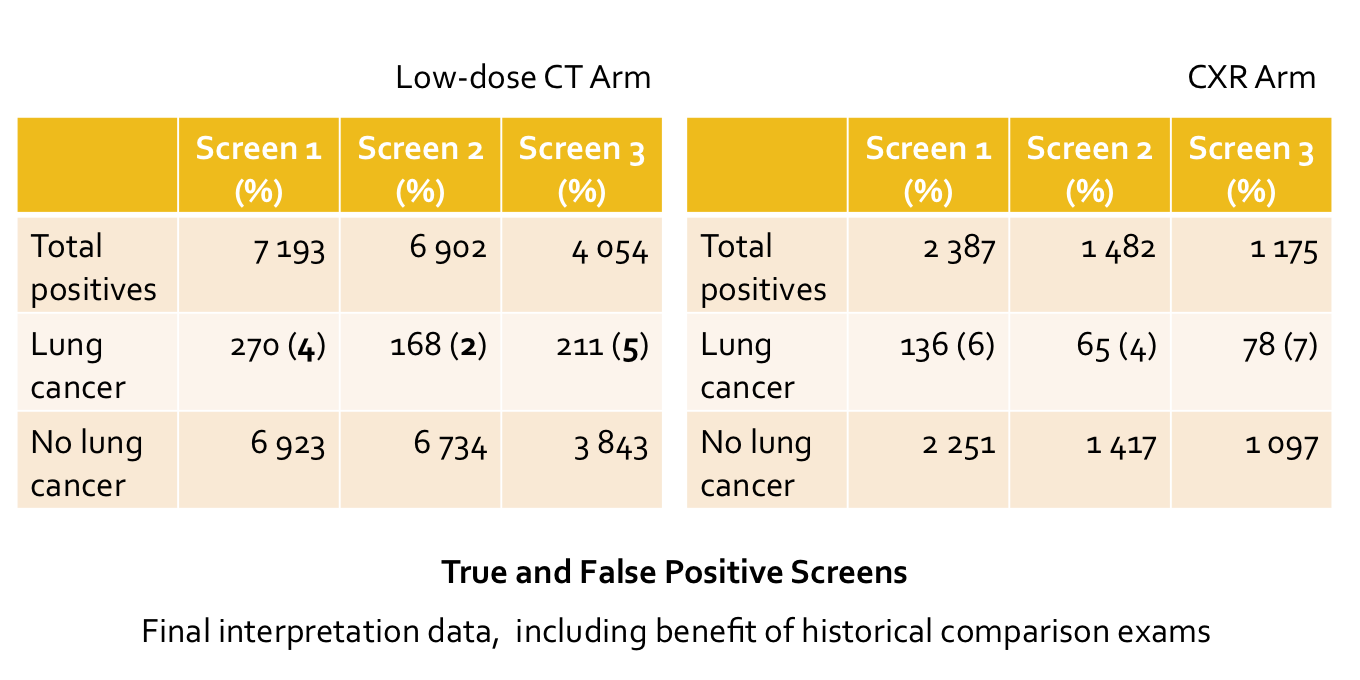
The tables show data from the NLST, with true and false positive screens at screening round #1, 2 and 3.

  • NLST is a randomized control trial studying the effect of low-dose CT screening vs. CXR in reduction of lung cancer-specific mortality. The interim result had been announced in October 2010, which showed a 20% reduction in mortality of those who had screened with low-dose CT.
  • Overall positivity rates (non-calcified nodule 4 mm or greater in size and other findings potentially related to lung cancer) for the NLST are 24.2% at low-dose CT, and 6.9% at CXR
  • Of all positives, only 2-5% (at CT) and 4-7% (at CXR) of cases are truly lung cancer.
  • Number of false positives are relatively high.
  • To reduce false positives, systematic and multidisciplinary approach in establishing the regimen for low-dose CT lung cancer screening is needed.
Reference:
Gierada DS. RSNA 2010

February 20, 2011

Pulmonary Embolism Caused by Acrylic Cement

An axial CT image shows hyperdense structures within the segmental branch of the right middle lobe pulmonary artery (arrow), and in the perivertebral venous plexus (arrowheads). The patient had recent vertebroplasty performed for lumbar back pain.

Facts: Vertebroplasty
  • Injection of acrylic cement (polymethylmethacrylate; PMMA) into the diseased vertebral body for partial remodeling and lumbar pain relief
  • Infrequent complications such as infection, cement leakage
Facts: Leakage from Vertebroplasty
  • Can be into the spinal canal, or perivertebral venous plexuses
  • Perivertebral venous leakage can be into the internal venous plexuses (potentially resulting in cord/nerve root compression) or external venous plexuses
  • More often if inadequate preparation of PMMA (acrylic cement must be mixed to consistency of paste before injection to ensure advanced polymerization)
  • More often if vertebroplasty is performed for vascular lesions (metastasis from thyroid cancer, renal cell carcinoma, vertebral angiomas)
Imaging Findings
  • Hyperdense material in the branches of pulmonary arteries with or without evidence of pulmonary infarction
  • Hyperdense materials in the perivertebral veins

Reference:

Padovani B, et al. Pulmonary embolism caused by acrylic cement: a rare complication of percutaneous vertebroplasty. AJNR 1999; 20:375-377.

ShareThis