
 Fig.1: Transverse ultrasound image at the region of the gallbladder (arrow) shows a nearby large cystic structure (star).

Fig. 2 and 3: Axial T2 MR image and MRCP confirms the presence of the large cystic lesion adjacent to the gallbladder (arrow), proven to be saccular dilatation of the extrahepatic bile duct.
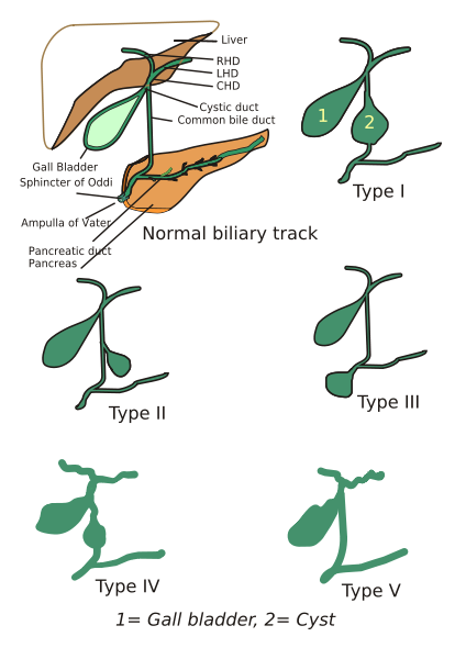
Fig.4: Diagram showing Todani classification of choledochal cyst
Choledochal Cyst
- Rare, congenital dilatation of biliary tree
- Can be either intra-hepatic, extra-hepatic or both
- Female:male ratio = 4:1, more common in Asia
- At risk for recurrent cholangitis, stricture, stone, pancreatitis and malignancy
- Risk of malignant transformation increases with age, and more often in type I and IV cysts.
Todani Classification
- Type I - fusiform dilatation of extrahepatic duct
- Type II - focal saccular dilatation or diverticulum of extrahepatic duct
- Type III - cystic dilatation of bile duct confined to duodenal wall (choledochocele)
- Type IVa - combined intra and extrahepatic duct dilatation
- Type IVb - multiple extrahepatic duct dilatation
- Type V - multiple intrahepatic biliary cysts (Caroli's disease)
Our case - Todani type II choledochal cyst
Image source: diagram from www.en.wikipedia.org/_wiki/Choledochal_cysts
Reference:
1. Wiseman K, Buczkowski AK, Chung SW, et al. Epidemiology, presentation, diagnosis, and outcomes of choledochal cysts in adults in an urban environment. Am J Surg 2005;189:527-531.


No comments:
Post a Comment