December 21, 2012

Atypical Hepatic Hemangioma - Rapid Filling of Contrast

Axial MR images in multiple phases (as labeled) show a T1 hypointense nodule in the right hepatic lobe that rapidly filled in with contrast after administration and subsequently fades on delayed imaging. Note that the enhancement of the nodule is similar to the aorta in all phases. On T2W image (not shown), this nodule has a very high signal intensity. 


Rapidly Filling Hemangioma
  • 16% of all hepatic hemangiomas
  • More common among small (<1 1="1" 42="42" cm="cm" hemangioma="hemangioma" hemangiomas="hemangiomas" in="in" incidence="incidence" less="less" li="li" of="of" than="than">
  • Immediate homogeneous enhancement at arterial phase and hyperintensity persists in delayed phases. Enhancement equal to aorta in all phases. 
  • High T2 signal intensity may be helpful for differentiation from other arterial enhancing nodules (but it can also be seen in islet cell tumor metastasis)

Reference:
Vilgrain V et al. Imaging of atypical hemangiomas of the liver with pathologic correlation. Radiographics 2000; 20:379

December 11, 2012

Pelvic Ewing's Sarcoma

A pelvic radiograph of a 5-year-old girl shows a large lytic lesion in the left iliac bone (arrows).
An axial FDG PET/CT image shows high metabolic activity of the mass involving the left iliac bone with soft tissue component and bone destruction. A coronal T2W MR image reveals an extensive soft tissue mass with necrotic areas and involvement of the adjacent musculature. 


Differential Diagnosis
  • Metastatic neuroblastoma. Given her age at five years old, this needs to be in differentials
  • Ewing's sarcoma
  • Telangiectatic osteosarcoma
  • Osteomyelitis. Great mimics of aggressive-looking bone tumor. Symptoms may overlap with round-cell tumor, including fever
This case: Ewing's sarcoma by tissue diagnosis (+ve PAS and vimentin).

Facts: Ewing's Sarcoma

  • Malignant round-cell tumors of the bone with neural cell origin
  • Tumors of children and young adults, most between 10-20 years old. Less than 2% occur in children less than 5 years old
  • Most common sites = femur >> pelvis
  • Pelvic Ewing's -- bad prognosis because there is no anatomic barrier to tumor spread, close proximity to viscera and neurovascular bundles, prone to recur

Reference:
Bhagat S, Sharma H, Pillai DS, Jane MJ. Pelvic Ewing's sarcoma: a review from Swedish Bone Tumour Registry. J Orthop Surg 2008;16:333-8

November 30, 2012

Maisonneuve Fracture

Figure 1: AP and lateral ankle radiographs demonstrate a vertical fracture of the medial/posterior malleolus of the distal tibia without a fibular fracture.  

Figure 2: Full-length AP fibular radiograph shows a mildly displaced fracture of the fibular shaft at the junction between the proximal 1/3 and middle 1/3. 

Facts: Rotational Ankle Fractures

  • Rotational ankle fractures are classified according to force direction applied to the foot, while the injured foot can be in a different position (supination/pronation, adduction/external rotation)
  • AO/Weber classification: A, B, C fractures are differentiated by location of fibular fractures. 
  • Fibular fracture below the syndesmosis = AO/Weber A (usually supination-adduction)
  • Fibular fracture at the syndesmosis = AO/Weber B (~ supination and external rotation)
  • Fibular fracture above the syndesmosis = AO/Weber C (~ pronation external rotation)
Facts: Maisonneuve Fractures
  • High fibular fracture above the syndesmosis resulting from external rotation
  • Often, there is injury to the medial ankle either a tranverse medial malleolar fracture, posterior malleolar fracture or disruption of the deltoid ligament
  • Disruption of the syndesmosis and interosseous ligament up to the fibular fracture site
  • Suspicious for this fracture if you see a 1) transverse medial malleolar fracture or 2) posterior malleolar fracture but no fibular fracture on the ankle radiographic series. In these situation, a full-length fibular radiograph should be taken

Reference:
Sakthivel-Wainford K. Self-assessment in limb x-ray interpretation, 2006
Rockwood CA, Green DP. Rockwood and Green's fractures in adults, 2005


November 26, 2012

Screening Mammography Overdiagnoses Breast Cancer?

The recently published NEJM's original article describes effect of screening mammography on breast cancer incidence in the USA. Bleyer A and Welch HG. N Eng J Med 2012;367:1998 (22 Nov 2012)


Facts: Aim of Cancer Screening

  • We expect cancer screening program to lower cancer-related mortality by a means of 1) earlier detection of disease destined to be fatal and 2) early Rx of screen-detected cancers
  • A cancer screening program should 1) increase incidence of cancer detected at an earlier stage and 2) decrease incidence of cancer presenting at a late stage

What Study Is About And What It Has Found:
  • Breast cancer rate from 1976 through 1978 (mammography was uncommon) ~ baseline incidence
  • Breast cancer rate from 2006 through 2008 ~ current incidence
  • Confounding effects of menopausal hormone therapy were minimized by not including transitory increase in incident breast cancers from 1990 through 2005. Underlying incidence of breast cancer assumed to rise by 0.25% annually
  • Number of cases of early-staged breast cancer rises from 112 to 234 cases per 100,000 women
  • Number of cases of late-staged breast cancer decreases from 102 to 94 cases per 100,000 women
  • "With an assumption of a constant underlying disease burden, only 8 of the 122 additional early-stage cancers diagnosed were expected to progress to advanced disease." This was estimated that breast cancer was overdiagnosed. In 2008, the overdiagnosis would account for 31% of all breast cancer diagnosed
Other Studies Said?
Results of this study concur with other reports (references 2-3) and suggest that improvements in treatment are the main drive in reducing breast cancer mortality - rather than screening mammography. 

It is suggested that "pros and cons of mammography should be incorporated into the counseling that women receive as they decide whether an when to be screened". 


References:
  1. Bleyer A and Welch HG. Effect of three decades of screening mammography on breast cancer incidence. N Eng J Med 2012;367:1998
  2. Kalager M et al. Effect of screening mammography on breast cancer mortality in Norway. N Eng J Med 2010;363:1203
  3. Autier P et al. Breast cancer mortality in neighbouring European countries with different levels of screening but similar access to treatment: trend analysis of WHO mortality database. BMJ 2011;343:d4411
  4. Kaunitz AM. Screening mammography: does overdiagnosis overshadow prevention of advanced breast cancer? Journal Watch Women's Health November 21, 2012

November 20, 2012

Spinal Langerhans Cell Histiocytosis (LCH)

Figure 1: Tc-99m MDP bone scan (posterior image) shows a focus of increased activity at L2 vertebral body in a 24-year-old woman presenting with back pain.

Figures 2&3: Axial GRE T2W and sagittal post-contrast T1W MR images show a round focus of bone destruction surrounded by bone marrow edema and enhancement of L2 body, sparing the posterior elements. 

Facts: LCH
  • Rare, benign disorder of unknown etiology comprises of eosinophilic granuloma (unifocal), Hand-Schuller-Christian disease (multifocal) and Letterer-Siwe disease (disseminated variant) - these are different manifestations of a same disease
  • Clonal proliferation of Langerhans cells
  • Peak incidence 5-10 years but there is a shift toward younger children
Spinal LCH
  • Vertebral involvement in 8-25% of cases
  • Thoracic > cervical, lumbar
  • Vertebral body >> posterior elements
  • Solitary, well-defined osteolytic lesion with scalloped borders eventually progresses to collapse and a classic "vertebra plana"
  • Typically single vertebral body involved. Disc spaces spared
  • Soft tissue mass suggests more aggressive course
Main Imaging Differentials of Spinal LCH
  • Osteomyelitis
  • Ewing sarcoma
  • Leukemia, lymphoma, metastatic neuroblastoma

Reference:
Hosalkar HS, Greenberg JS, Wells L, Dormans JP. Isolated Langerhans Cell Histiocytosis of the T12 vertebra in an adolescent. Am J Orthop 2007;36: E21-E24.

November 10, 2012

Parotid Warthin Tumor


Figures 1&2: Coronal and sagittal-reformatted CT images of an 80-year-old woman show a well circumscribed mass in the tail of the right parotid gland. There is homogeneous enhancement. 

Facts:  Warthin Tumor
  • Also known as papillary cystadenoma lymphomatosum
  • 14-30% of all parotid tumors
  • Almost always in older adults (peak incidence at 6th-7th decades of life)
  • Most commonly found within parotid gland (esp. tail). Occasionally in minor salivary glands.
  • MRI, although has high specificity and sensitivity for diagnosis, still cannot differentiate all malignant parotid tumors from benign ones. Tissue sampling is required for definitive diagnosis
Imaging:
  • Well-circumscribed solid mass, homogeneous enhancement and without calcification
  • If calcification is present in a benign-appearing parotid mass, pleomorphic adenoma should be considered first
  • US: multiple anechoic internal areas
  • MRI: low T1, high T2 signal intensity
  • Salivary scintigraphy: Accumulation of Tc-99m pertechnetate due to high mitochondrium

Reference:
Hatch RL, Shah S. Warthin tumor: a common, benign tumor presenting as a highly suspicious mass. J Am Board Fam Med 2005;18: 320-322.

November 1, 2012

Focal Fat Sparing

Figure 1: US image of the liver shows focal masslike area of hypoechogenicity of the left lobe posterior to the left portal vein branch. Note high echogenicity of the background liver, suggesting fatty change.

Figure 2 & 3: In-phase and out-of-phase MR images show liver signal intensity drop in the chemical shift imaging confirming diffuse fatty liver. The abnormality in the left lobe liver does not change between the two phases, suggesting a focal area of fat sparing.

Facts: Fatty Liver
  • Most common abnormality of the liver seen on cross-sectional imaging
  • Common patterns: diffuse fat accumulation, diffuse fat accumulation with focal sparing, and focal fat accumulation in an otherwise normal liver
  • Unusual patterns may mimick neoplasm, inflammation or vascular conditions
  • Pathology: triglyceride acculation within cytoplasm of hepatocytes
  • Term "fatty liver" is preferred over "fatty infiltration of the liver" because triglyceride accumulation occurs within hepatocytes but rarely other cell types. Infiltration of fat into parenchymal does not occur
Imaging Findings and Sensitivity/Specificity
  • US: 1) Liver echo greater than renal cortex and spleen with attenuation of sound wave, 2) loss of definition of diaphragm, 3) poor delineation of intrahepatic architecture (to avoid false-positive diagnosis, all three findings should be fulfilled).  Sensitivity 60-100%. Specificity 77-95%.
  • CT: Liver attenuation 10 HU less than that of spleen, or less than 40 HU. Sensitivity 43-95%. Specificity 90%.
  • MRI: Signal intensity loss on opposed-phase images in comparison with in-phase images. Sensitivity 81%. Specificity 100%.
Patterns
  1. Diffuse deposition: most common
  2. Focal deposition and focal sparing: characteristically in specific areas (adjacent to falciform ligament or ligamentum venosum, porta hepatis, in GB fossa). Suggestive findings of fatty pseudolesions rather than true masses are:
    1. Fat content
    2. Characteristic location
    3. Absence of mass effect on vessels and other liver structures
    4. Geographic configuration (not round or oval)
    5. Poorly delineated margin
    6. Contrast enhancement similar to or less than that or normal liver parenchyma
  3. Multifocal deposition
  4. Perivascular deposition
  5. Subcapsular deposition

Reference:


Hamer OW, Aguirre DA, Casola G, et al. Fatty liver: imaging patterns and pitfalls. Radiographics 2006;26: 1637-1653.

August 31, 2012

ESWL-induced Perinephric Hematoma

Ultrasound image of the kidney shows a crescentic heterogeneous hypoechoic lesion in the perinephric space (arrows). The kidney is marked by the calipers. 
Axial unenhanced CT images confirm a thick left perinephric hematoma (stars) and several fragmented stones in the lower pole of the left kidney.
Facts
  • ESWL (Extracorporeal Shock Wave Lithotrypsy) is a common and standard treatment for renal/proximal ureteric calculi in majority of patients
  • Most common complication = microscopic hematuria
  • Perinephric hematoma and infection (including pyelonephritis) can occur
  • Incidence of hematoma varies depending on method of diagnosis. By US, incidence is about 0.1-0.6%. By CT/MRI, incidence rises to 20-25% of cases. 
  • No clear correlation between number of shockwaves or intensity given and incidence of hematoma
  • Most perinephric hematoma resolves spontaneously within 2 years and the renal function is preserved. They are mostly treated conservatively
Imaging: US and CT
  • Crescent-shaped collection surrounding the affected kidney
  • Hypoechoic on US, hyperattenuating on non-contrast CT and no enhancement after IV contrast
  • Displacement or compression of adjacent renal parenchyma
  • Differentiate from subcapsular hematoma by appearance and pressure effect to underlying kidney.
  • "Page" kidney occurs when (usually) subcapsular hematoma causes chronic renal parenchymal compromise and then hypertension

Reference:
Labanaris AP, Kuhn R, Schott GE, Zugor V. Perirenal hematomas induced by extracorporeal shock wave lithotripsy (ESWL). Therapeutic management. TheScientificWorldJOURNAL 2007;7:1563-1566.

August 21, 2012

Mediastinal Neuroblastoma

Axial CT image at the level of mid thoracic spine of a 5-year-old boy shows a well-circumscribed, enhancing left paraspinal mass located between the medial ends of the ribs. 

An MIBG study (posterior view of the thorax and upper abdomen) shows a focus of moderate uptake in the left side of the lower thorax, corresponding with the site of abnormal mass on CT. Note intense uptake of bilateral adrenal glands. 


Facts: Neuroblastoma

  • 8-10% of all childhood malignancies
  • Malignant tumor composed of immature ganglion cells. Most (2/3) arise from abdomen or pelvis and the rest is extra abdominal
  • Typically seen in children under age of 5 years
  • Treatment determined by stage of tumor at presentation. Regionally limited disease is potentially resectable, but locally extensive or disseminated disease is usually not resectable
  • Current imaging staging evaluation: 1) CT or MRI of primary tumor, 2) skeletal survey, 3) bone scintigraphy or MIBG for bone metastasis, and 4) bone marrow aspiration and biopsy for marrow disease
  • CT alone has low sensitivity (43%) but high specificity (97%) for detection of stage 4 disease. CT accuracy = 81%. MRI is more sensitive (83%) but less specific (88%) and slightly more accurate (85%). 
Imaging Findings: CT/MRI
  • Sharply marginated, fusiform paraspinal mass
  • Oriented along direction of sympathetic chain
  • 40% contain calcifications
  • Heterogeneous enhancement
  • Differentiation from ganglioneuroblastoma (usually 5-10 years old) and ganglioneuroma (usually > 10 years old) not possible by imaging. Basically, they are tumors of sympathetic ganglia


References
1. Siegel MJ. Pediatric Body CT. 2008
2. Siegel MJ, Ishwaran H, Fletcher BD, et al. Staging of neuroblastoma at imaging: report of the radiology diagnostic oncology group. Radiology 2002;223:168-175. 

August 11, 2012

Avian Spur


Facts:


  • AKA supracondylar process of the humerus
  • Congenital osseous/cartilagenous projection arising from the anteromedial surface of the distal humerus
  • Found in 1% of population
  • Associated with ligament of Struthers, which connects the process to the medial epicondyle (fibers of pronator teres may arise from this structure)
  • Median nerve and brachial artery pass below this arch and may be compressed
  • Fracture is possible but rare
Reference:
Egol KA, Koval KJ, Zuckerman JD. Handbook of fractures; 4th ed, 2010. 

August 1, 2012

Superficial Femoral Vein: Misleading Medical Nomenclature

Anatomy and Definition of Superficial Femoral Vein (SFV)
  • SFV, as understood by vascular surgeons and radiologists, is a continuation of the popliteal vein. After joining the deep femoral vein, it becomes common femoral vein
  • Superficial femoral vein is actually a "deep" vein
The Problem
  • Most vascular surgeons and radiologists understand that SFV is a deep vein, but many physicians in other specialty or general practitioners do not
  • Based on a survey of multispecialty groups, only 24% of physicians would give anticoagulants to patients having "acute thrombosis of the superficial femoral vein". There is a misperception of many physicians that SFV is superficial vein, therefore it would not be treated as deep vein thrombosis
Recommendations: Don't Use "Superficial Femoral Vein". Use "Femoral Vein"
  • Current consensus developed by experts in phlebology officially established “femoral vein” as the vein that originates from the popliteal vein and courses in the femoral canal and bluntly discarded “superficial femoral vein” as an “unauthorized term" … because it is a deep vein 
  • SFV is not in the official Terminologica Anatomica
  • The other vein is "deep femoral vein" or "profunda femoris vein"
  • Supported by International Interdisciplinary Consensus Committee on Venous Anatomical Terminology convened on September 8–9, 2001 (Nomenclature of the veins of the lower limbs: an international interdisciplinary consensus statement. J Vasc Surg 2002; 36:416-422)
  •  Supported by Society of Interventional Radiology

Reference:
Hammond I. The superficial femoral vein. Radiology 2003;229;604-666 (link)

July 21, 2012

Pyonephrosis

 Longitudinal ultrasound image of the right kidney shows moderate right hydroureteronephrosis (arrows) with internal debris.

Longitudinal image of the right ureter (arrows) shows a stone (arrowhead) in the distal ureter causing proximal hydroureter. 

Facts:

  • Bacterial infection of urine associated with ureteral obstruction, AKA infected hydronephrosis. Accumulation of pus in the renal pelvis and calices of the kidney
  • Common causes are ureteric obstruction by stone and ureteropelvic junction (UPJ) obstruction
  • Septic patients with high fever, flank pain and tenderness
  • Any febrile patients with hydronephrosis should be suspected of having pyonephrosis
  • Ultrasound: echogenic urine and debris in the hydronephrotic kidney
  • Prompt drainage essential 
Reference:
Hodler J, Von Schulthess GK, Zollikofer ChL. Diseases of the abdomen and pelvis 2010-2013

July 12, 2012

"Thai Radiology News" Image Quiz & Answer 1/2555

Author: พ.ญ.​ นิธิมา ศักดิ์โสภาวิวัฒน์  คณะแพทยศาสตร์โรงพยาบาลรามาธิบดี
Editor: น.พ. รัฐชัย แก้วลาย คณะแพทยศาสตร์โรงพยาบาลรามาธิบดี

ประวัติ:
ผู้ป่วยชาย อายุ 27 ปีมาตรวจที่แผนกฉุกเฉินด้วยอาการเจ็บคอด้านขวา ร่วมกับอาการกลืนเจ็บ กลืนลำบาก เป็นมาประมาณ 10 วัน ต่อมามีไข้ และหายใจลำบาก


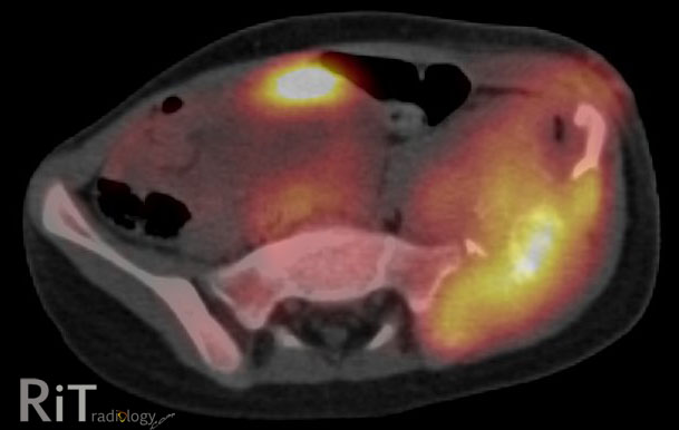
ภาพที่ 1 เป็น axial CT ส่วนคอแสดงให้เห็น rim-enhancing fluid collection ทางด้านขวา หน้าต่อ carotid space. ภายใน carotid space ไม่เห็น internal jugular vein ที่ปกติ แต่ถูกแทนที่ด้วย hypodense filling defect ร่วมกับผนังของหลอดเลือดดำมี enhancement. ภาพที่ 2 เป็นภาพ coronal reformation เพ่ิมเติม แสดงให้เห็น filling defect ภายใน internal jugular vein ข้างขวาและ enhancement ของผนังหลอดเลือดนี้. ภาพที่ 3 เป็นภาพ axial CT ทรวงอกใน lung window แสดงให้เห็น pulmonary nodules ขนาดเล็กกว่าหนึ่งเซนติเมตรในปอดทั้งสองข้าง บางอันมี cavity ภายใน สังเกตว่า nodules มักอยู่ที่รอบนอกของปอด (Image courtesy of University of Maryland Medical Center, MD, USA)

การวินิจฉัยโรค            Lemierre Syndrome

Discussion             ผู้ป่วยรายนี้มาพบแพทย์เนื่องจากมีการอักเสบติดเชื้อที่ต่อมทอนซิลและเกิด peritonsillar abscess ทำให้มี thrombophlebitis ของ internal jugular vein ที่อยู่ติดกันและมี septic emboli กระจายไปยังปอดทั้งสองข้าง  โรคติดเชื้อในช่องปากและคอที่มีภาวะแทรกซ้อนเช่นนี้รู้จักกันในชื่อ Lemierre syndrome ซึ่งตั้งตามชื่อแพทย์ผู้รายงานกรณีผู้ป่วย 20 ราย ในปี พ.ศ. 2479 (1) ในปัจจุบันพบโรคนี้น้อยมากเนื่องจากมีการใช้ยาปฏิชีวนะกันอย่างแพร่หลาย แต่การวินิจฉัยภาวะนี้ให้รวดเร็ว แม่นยำ ยังมีความสำคัญเพราะหากได้รับการรักษาช้าอาจทำให้เสียชีวิตได้
Lemierre syndrome ส่วนใหญ่เกิดจากการติดเชื้อ anaerobic Fusobacterium necrophorum เมื่อมีการติดเชื้อที่ช่องคอ ทอนซิลหรือช่องปากแล้วอาจมีการแพร่กระจายโดยตรง, ผ่านทางหลอดเลือดรอบๆต่อมทอนซิลหรือผ่านทางท่อน้ำเหลืองไปยัง internal jugular vein ทำให้เกิด thrombophlebitis และ septic emboli กระจายไปยังอวัยวะอื่นๆ ได้  ที่พบบ่อยที่สุดคือปอด (79-100%) ผู้ป่วยส่วนมากเป็นวัยรุ่นแต่ก็พบได้ทุกอายุ มักมาพบแพทย์ด้วยอาการเจ็บคอ มีไข้ บวมกดเจ็บ และอาการอื่นๆ ขึ้นกับตำแหน่งการแพร่กระจายของ septic emboli (2, 3).
การตรวจทาง imaging มีบทบาทมากในการวินิจฉัยภาวะนี้ อัลตราซาวด์สามารถแสดงให้เห็นลิ่มเลือดที่อยู่ภายใน internal jugular vein ได้ดี ไม่ว่าจะเป็นชนิดที่มีการอุดกั้นหรือไม่ก็ตาม หรืออาจเห็นลักษณะ venous distension, absent flow หรือ non-compressible vein. การตรวจด้วย CT ร่วมกับการให้สารทึบรังสีชนิดฉีดจะแสดงขอบเขตของการติดเชื้อ เช่น peritonsillar abscess ได้ดีกว่าอัลตราซาวด์ และแสดงภาพลิ่มเลือดเป็น filling defect ภายในหลอดเลือดดำ  หลอดเลือดดำดังกล่าวอาจมีขนาดใหญ่ขึ้นร่วมกับมี fat stranding รอบๆ และมี enhancement ของผนังหลอดเลือด. การตรวจเอกซเรย์ปอดอาจพบว่ามี pulmonary nodules, masses, cavitary lesions, focal airspace disease ได้แต่การทำเอกซเรย์คอมพิวเตอร์จะให้รายละเอียดได้ดีกว่า ความผิดปกติที่พบในปอดมักอยู่ที่บริเวณรอบนอก (periphery) ของเนื้อปอด เข้าได้กับ septic emboli  หากสงสัยภาวะนี้ ผู้ป่วยควรได้รับการตรวจ CT ของทรวงอกและคอในคราวเดียวกัน  การรักษาได้แก่การให้ยาปฏิชีวนะ ยาละลายลิ่มเลือด และการผ่าตัด (4).

เอกสารอ้างอิง
1.            Lemierre A. On certain septicemias due to anaerobic organisms. Lancet 1936; 1:701-703.
2.            Karkos PD, Asrani S, Karkos CD, et al. Lemierre's syndrome: A systematic review. Laryngoscope 2009; 119:1552-1559.
3.            Vargiami EG, Farmaki E, Tasiopoulou D, et al. The Lemierre syndrome. Eur J Pediatr 2010; 169:411-414.
4.            Weeks DF, Katz DS, Saxon P, Kubal WS. Lemierre syndrome: report of five new cases and literature review. Emerg Radiol 2010; 17:323-328.

July 11, 2012

Miliary Tuberculosis

A chest radiograph of a 37-year-old woman shows innumerable tiny nodules throughout both lungs in a uniform, symmetric distribution. 

Axial chest CT image confirms the presence of innumerable micronodules in a random distribution and uniformity.

Facts: Miliary TB

  • Acute disseminated infection of mycobacterium tuberculosis via bloodstream, or progression of active TB (rupture of caseating lymph node or cavity into blood vessel)
  • Primary site may be intra- or extrapulmonary, or may not be recognizable
  • Life-threatening infection. Mortality 13-50%
  • Any age, any immune status but poor immune individuals are at increased risk
  • Only 30% have positive sputum examination
  • Up to 60% have negative skin test
Imaging
  • Chest radiograph (CXR) can be normal in early disease 
  • CXR usually shows abnormality up to 10 days or more after clinical illness has started. CT can show it earlier
  • "Miliary" nodules are tiny (<2 mm), discrete, about the same size. They may coalesce into patchy and more irregular opacities
  • Nodules may take 2-3 months to fade even with adequate therapy
  • No correlation between number or size of nodules and clinical health
  • Lymph node enlargement, hepatosplenomegaly may be seen
Reference:
Palmer PES, Wambani SJ, Reeve P. The imaging of tuberculosis: with epidemiological, pathological, and clinical correlation, 2001. 

July 1, 2012

Avulsion of the Anterior Superior Iliac Spine

A pelvic radiograph demonstrates an avulsion fracture (arrows) of the right anterior superior iliac spine (ASIS) in a 14-year-old boy. 

Facts: Pelvic Avulsions

  • Avulsion of pelvic bones usually found in young, skeletally immature athletes.
  • Forceful contraction of the attached muscle while the athlete actively engages in kicking, running or jumping.
  • Three major locations: ASIS (sartorius attachment), anterior inferior iliac spine (AIIS, rectus femoral attachment) and ischial tuberosity (hamstrings and adductor attachment).
  • 50% of cases at ischial tuberosity, 23% ASIS, 22% AIIS (of all pelvic avulsions).
  • Localized swelling and tenderness at the site of avulsion fracture. Limited motion from pain.
Imaging
  • Plain radiography usually sufficient for diagnosis. 
  • Comparison view helpful to ensure that abnormality is not a secondary center of ossification.
  • Pitfalls: secondary ossification center, osseous mass seen as a delayed presentation mimicking neoplasm.
References
Davies AM, Johnson KJ, Whitehouse RW. Imaging of the hip & bony pelvis: techniques and applications. 
Beaty JH, Rockwood CA, Kasser R. Rockwood and Wilkins' fractures in children. 

May 31, 2012

Acute Isodense Subdural Hematoma


Plain and contrast-enhanced axial CT images of the brain show an isodense subdural hematoma (SDH, arrows) in the left cerebral convexity, much better appreciated on post-contrast image. There is also a thinner right frontal convexity SDH. 


Facts: Isodense Subdural Hematoma (SDH)
  • Subdural blood collection that has similar attenuation with the gray matter
  • Acute SDH appears as a high density collection with declining density with time. It passes "isodense" state mostly in subacute phase (2-6 weeks after initial trauma)
  • Isodense SDH poses diagnostic dilemma because it is not apparently seen on CT
  • In acute setting this can be seen in anemic patients (acute isodense SDH). Experimental data showed that Hb 8-10 g/dl will be isodense to the adjacent brain

Reference:
Smith, Jr., WP, Batnitzky S, Rengachary SS. Acute isodense subdural hematomas: a problem in anemic patients. AJR 1981; 136:543-546. 

May 21, 2012

USPSTF Recommends Against PSA-based Prostate Cancer Screening


In its newest Statement published yesterday in the Annals of Internal Medicine, the U.S. Preventive Services Task Force (USPSTF) recommends against PSA-based screening for prostate cancer. Read the full paper (free) here.

Prostate Cancer: Facts
  • Most commonly diagnosed non-skin cancer in men in USA, estimated lifetime risk 15.9%
  • Most cases have good prognosis even without treatment
  • Lifetime risk of dying of prostate cancer 2.8%
Screening with PSA
  • PSA-based screening programs detect many cases of asymptomatic prostate cancer but evidence suggests that many of them will not progress or will progress slowly that it would have remained asymptomatic for the lifetime
  • "Overdiagnosis" of prostate cancer based on PSA is between 17-50% 
  • Screening resulted in none or minimal reduction in prostate cancer mortality (0 to 1 prostate cancer deaths avoided per 1000 men screened)
  • "False positivity" near 80% (cutoffs value 2.5-4 ug/L)
Recommendation
  • Applies to men in general US population. Although older age is the strongest risk factor for development of prostate cancer, neither screening nor treatment trials show benefit in men older than 70 years
  • Decision to initiate or continue PSA screening should be understood by patients about possible benefits and harms of screening
Recommendations of Others
  • The American Urological Association, the American Academy of Family Physicians and the American College of Physicians: currently updating their guideline
  • The American Cancer Society: men at average risk beginning at age 50 years and black men or men with a family history of prostate cancer beginning at age 45 years
Reference:
Annals of Internal Medicine May 21, 2012  LINK

May 11, 2012

ACR Appropriateness Criteria for Suspected Aortic Injury

Axial contrast-enhanced CT image shows a pseudoaneurysm (arrow), intimal flap and periaortic hematoma of the proximal descending thoracic aorta in a patient experienced severe blunt chest trauma.

A newly revised American College of Radiology (ACR)'s Appropriateness Criteria for blunt chest trauma - suspected aortic injury has been published in March 2012 in the journal Emergency Radiology, summary and useful points are provided below

  • Chest x-ray remains an initial screening examination in patients who has sustained blunt chest trauma
  • In the appropriate clinical setting and with a CXR demonstrating mediastinal widening or other signs of mediastinal hemorrhage, thoracic aortography or helical chest CT is indicated
  • CTA is emerging as a very sensitive and specific examination for aortic injury and has replaced aortography in many trauma centers
Useful Points
  • Mediastinal widening has been defined as a transverse diameter of 8 cm from the left side of aortic arch to the right margin of the mediastinum (even on AP portable CXR)
  • Mediastinal widening is 90% sensitive but 10% specific for aortic injury
  • Approx 7% of patients with aortic injury have normal initial CXR
  • If no mediastinal hematoma seen on CT, probability of significant aortic injury is very low
Reference
Demehri S, et al. ACR Appropriateness Criteria blunt chest trauma--suspected aortic injury. Emerg Radiol 2012 (published online: 18 Mar 2012)

May 1, 2012

Age-related White Matter Changes


MR FLAIR images show multiple FLAIR hyperintense foci in bilateral periventricular and deep white matter in a 77-year-old patient.

Facts:
  • Several possible mechanisms but at present it is believed to be an incomplete ischemia mainly related to cerebral small vessel arteriolosclerosis
  • Pathology: partial loss of myelin, axons, oligodendroglial cells; mild reactive gliosis, astrocytic gliosis, stenosis of arterioles from hyaline fibrosis
  • Important substrates for cognitive impairment and functional loss in the elderly
  • Very common in elderly with hypertension, diabetes, hyperlipidemai
Imaging
  • Ill-defined hypodensities on CT
  • T1 hypointensities, T2/PD/FLAIR hyperintensities. FLAIR best to demonstrate severity of disease
  • When the largest lesion is adjacent to ventricles, it's called periventricular white matter changes
  • On diffusion tensor MRI, there is elevation of diffusivity and reduced fractional anisotrophy (FA) meaning impaired white matter integrity

Reference:
Xiong YY, Mok V. Age-related white matter changes. J Aging Res 2011

April 21, 2012

Sternal Foramen

Axial, sagittal-reformatted and 3D CT images of the sternum shows a small round defect in the lower third of the sternum (arrows)

Facts: Sternal Foramen
  • Small round/oval defect in the lower third of the sternum
  • Incomplete fusion of multiple ossification centers
  • Usually asymptomatic and of no clinical significance
  • Prevalence 6.7% in autopsy population
Imaging
  • Mostly incidentally found on chest CT
  • Usually solitary
  • Mostly in the body of the sternum. Rare in the manubrium

Reference:
Yekeler E, et al. Frequency of sternal variations and anomalies evaluated by MDCT. AJR 2006;186:956-960


April 10, 2012

Osteoid Osteoma

A frontal view of the right femur of a teenage boy demonstrates an ill-defined sclerotic area (arrows) in the proximal diaphysis with thickened cortex.

A coronal-reformatted CT image shows a well-defined lucency (arrow) within the central portion of sclerotic medulla. Within that lucency, a tiny calcific nidus is seen. Thickened cortex is also observed.

Facts: Osteoid Osteoma
  • Self-limited benign osteogenic tumor consisting of a vascular mass (nidus) surrounded by reactive bone sclerosis
  • Male predominance (male:female = 2:1). Teenagers and young adults (90% of cases between 5-30 years old)
  • Characteristic pain referring to the nearest joint, worse at night. Pain is relieved by aspirin or NSAIDs
  • Treatment options: surgical excision, CT-guided percutaneous resection or destruction of the nidus
Imaging Appearance
  • Location: cortex, medulla or periosteum (anywhere but cortex most common)
  • Long bones of lower extremity (esp femoral neck) most commonly affected. Almost never seen in flat bones and craniofacial bones
  • Central lucent area (nidus) surrounded by sclerotic bone (nidus may be subtle and has variable degree of calcification)
  • CT is helpful to identify the nidus (as in our case)
  • MRI can be misleading because reactive bone marrow edema and soft tissue involvement may mimic malignancy
Our case: osteoid osteoma centered in the medullary cavity. The diagnosis was confirmed by CT (showing a lucent nidus with calcification) and clinical picture.

Reference:
Kadir S. Teaching Atlas of Interventional Radiology, 2005.
Vioria VJ, et al. Orthopaedic Pathology,

ShareThis