December 21, 2012

Atypical Hepatic Hemangioma - Rapid Filling of Contrast

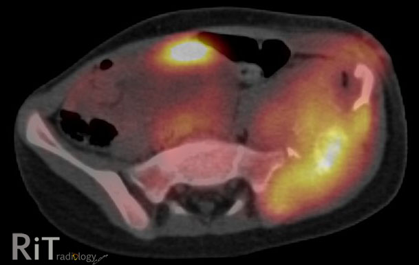
Axial MR images in multiple phases (as labeled) show a T1 hypointense nodule in the right hepatic lobe that rapidly filled in with contrast after administration and subsequently fades on delayed imaging. Note that the enhancement of the nodule is similar to the aorta in all phases. On T2W image (not shown), this nodule has a very high signal intensity. 


Rapidly Filling Hemangioma
  • 16% of all hepatic hemangiomas
  • More common among small (<1 1="1" 42="42" cm="cm" hemangioma="hemangioma" hemangiomas="hemangiomas" in="in" incidence="incidence" less="less" li="li" of="of" than="than">
  • Immediate homogeneous enhancement at arterial phase and hyperintensity persists in delayed phases. Enhancement equal to aorta in all phases. 
  • High T2 signal intensity may be helpful for differentiation from other arterial enhancing nodules (but it can also be seen in islet cell tumor metastasis)

Reference:
Vilgrain V et al. Imaging of atypical hemangiomas of the liver with pathologic correlation. Radiographics 2000; 20:379

December 11, 2012

Pelvic Ewing's Sarcoma

A pelvic radiograph of a 5-year-old girl shows a large lytic lesion in the left iliac bone (arrows).
An axial FDG PET/CT image shows high metabolic activity of the mass involving the left iliac bone with soft tissue component and bone destruction. A coronal T2W MR image reveals an extensive soft tissue mass with necrotic areas and involvement of the adjacent musculature. 


Differential Diagnosis
  • Metastatic neuroblastoma. Given her age at five years old, this needs to be in differentials
  • Ewing's sarcoma
  • Telangiectatic osteosarcoma
  • Osteomyelitis. Great mimics of aggressive-looking bone tumor. Symptoms may overlap with round-cell tumor, including fever
This case: Ewing's sarcoma by tissue diagnosis (+ve PAS and vimentin).

Facts: Ewing's Sarcoma

  • Malignant round-cell tumors of the bone with neural cell origin
  • Tumors of children and young adults, most between 10-20 years old. Less than 2% occur in children less than 5 years old
  • Most common sites = femur >> pelvis
  • Pelvic Ewing's -- bad prognosis because there is no anatomic barrier to tumor spread, close proximity to viscera and neurovascular bundles, prone to recur

Reference:
Bhagat S, Sharma H, Pillai DS, Jane MJ. Pelvic Ewing's sarcoma: a review from Swedish Bone Tumour Registry. J Orthop Surg 2008;16:333-8

ShareThis