Showing posts with label MSK. Show all posts

April 21, 2014

Metatarsal Stress Fracture

Oblique radiographic view of the foot shows transverse fracture lines of the proximal diaphyses of the forth and fifth metatarsals (arrows). Note sclerotic bone ends, periosteal reaction and minimal widening of the fracture gaps (degree of sclerosis is more on the forth digit)
Facts

  • Spontaneous fractures of normal bone that result from summation of stresses
  • Most common lower-extremity stress fracture
  • Originally termed "march fracture" (seen in military recruits). Now seen in ballet, football, gymnastics and basketball
  • Most common site = shaft (at diaphysis or neck)
  • Increased incidence in pes cavus and pes planus foot


Radiography

  • Often negative in early phase. May see thickening of cortex and small periosteal reaction
  • Later, a fracture line with sclerotic bone ends, periosteal reaction, widening of fracture gap will be shown. 
  • Late phase, the bone ends involved are entirely sclerotic 

References:
Schepsis AA, Busconi BD. Sports Medicine, 2006.
Baxter DE, Porter DA, Schon L. Baxter's the Foot and Ankle in Sport, 2008. 

March 11, 2014

Tibial Spine Fracture in Adults

A lateral knee radiograph of a 22-year-old man sustaining motor vehicle collision demonstrates an oval bone fragment (arrow) in the intercondylar region of the knee. There is complete separation between the fragment and the donor site with superior displacement of the fragment. Note hemarthrosis (asterisk).

Facts:

  • Classically described in pediatric patients and considered the childhood equivalent of anterior cruciate ligament (ACL) ruptures in adults
  • Forceful hyperextension of the knee resulting in avulsive force/tension on ACL, which inserts into the anterior tibial spine. Possibly with valgus stress or rotation. 
  • In adults, most injuries occur in road-traffic accidents and are isolated
  • Adults more likely to have associated tear of medial collateral ligament (MCL) or intra-articular fracture
Classification (Meyers and McKeever)
  • Based on degree of displacement. Type II & III are most common
  • Type I = incomplete avulsion of tibial spine without displacement
  • Type II = incomplete avulsion with anterior elevation of the fragment
  • Type IIIA = complete separation of fragment
  • Type IIIB = rotated and comminuted fragment
  • Generally, types I and II are managed conservatively while type III fractures are managed arthroscopically or with open reduction

References:
Kendall NS, et al. Fracture of the tibial spine in adults and children. J Bone J Surg [Br] 1992;74-B:848-52.
Rosen's Emergency Medicine - Concepts and Clinical Practice

August 30, 2013

Medial Epicondyle Fracture of the Humerus

AP views of both elbows of an 18-year-old boy who sustained an injury to the right elbow.  There is an avulsion fracture (arrow) of the medial epicondyle of the right humerus. Radiograph of the left side demonstrates different areas of distal humeral structures from medial to lateral: medial epicondyle, trochlea, capitellum and lateral epicondyle. 

Facts:

  • Common pediatric elbow fracture (3rd common, after supracondylar and lateral condylar fractures)
  • Valgus strain at elbow joint
  • Two main types: simple avulsion (1/2) and fracture-dislocation (occurring with lateral elbow dislocation; 1/2)
  • Indications for surgery include 1) displaced fragment trapped in joint preventing reduction, 2) ulnar neuropathy, 3) valgus instability, 4) open fracture
Imaging:
  • Look for displaced fragment trapped in the joint and degree of displacement because they might indicate surgery
  • In patients less than 8 years, trochlea may be non-ossified and this may be confused with fracture of medial condyle, which is rarer and could be more complicated
  • Another imaging Ddx is osteochondrosis
References:
Wilson JN. The treatment of fractures of the medial epicondyle of the humerus. J Bone J Surg 1960;42:778.
Gottschalk HP, Eisner E, Hosalkar HS. Medial epicondyle fracture sin the pediatric population. J Am Acad Orthop Surgeons 2012; 20:223.
Wheeless' Textbook of Orthopedics link

March 21, 2013

Sternal Osteomyelitis


Chest radiograph of a patient who had sternal pain, fever and discharge shows no obvious bony abnormality.


Sagittal STIR and coronal T1W MR images demonstrate bone marrow edema with soft tissue changes in the sternum and right sternoclavicular joint (arrows). 

Facts:

  • Uncommon infection of the sternum and sternoclavicular joint
  • Usually affecting drug addicts, individuals with history of recent subclavian catheter placement, and patients with chronic debilitating illnesses
  • Inciting organisms vary widely depending on demographics
  • High failure rates of medical treatment alone. Typical treatment includes surgical debridement and en bloc removal
Imaging
  • Radiograph is rarely helpful
  • CT may show bone destruction but this may be late because damage begins in the joint. Surrounding soft tissue abnormalities are often a useful sign.
  • MRI much more sensitive to detect joint and bone changes that are similar in findings to other areas of bone/joint infection
Reference: 
Shields TW et al. General Thoracic Surgery, 7th edition, 2009.

January 11, 2013

Extraosseous Myeloma


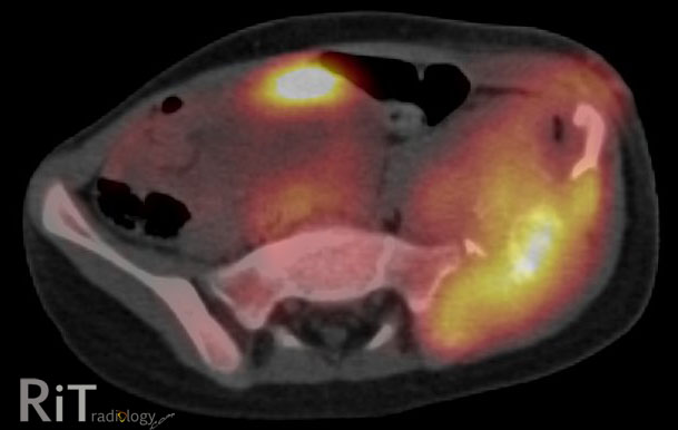
Axial PET/CT images show a large homogeneous soft tissue mass in the lett buttock involving the gluteal muscles, which demonstrates significant FDG avidity. 


Extraosseous Myeloma

  • 10-16% of patients with multiple myeloma
  • Rising incidence possibly due to better imaging techniques and increased survival of patients with myeloma
  • More common in younger myeloma patients and more aggressive subtypes
  • Shorter survival and shorter progression-free survival (than patients without extraosseous involvement)
  • Has been integrated into an updated version of Durie-Salmon staging system

How to Image Patients with Myeloma

  • Skeletal survey (current minimum standard)
  • Whole-body PET/CT vs. whole-body MRI for newly diagnosed myeloma and with no findings or limited findings on skeletal survey (two different methods have been suggested by different authorities) to assess for occult involvement
Imaging Appearances
  • Soft tissue: nodules in subcutaneous tissues and muscles (like in our case)
  • Reticuloendothelial system: lymph node enlargement in multiple stations
  • Liver: hepatomegaly, low-attenuation liver lesions without enhancement
  • CNS: leptomengineal process 
  • Lungs: lung nodules, masses, interstitial infiltration
  • Kidneys: masses, diffuse enlargement
  • Peritoneum: masses
Reference:
Hall MN, Jagannathan JP, Ramaiya NH, et al. Imaging of extraosseous myeloma: CT, PET/CT and MRI features. AJR 2010; 195:1057-1065. 

December 11, 2012

Pelvic Ewing's Sarcoma

A pelvic radiograph of a 5-year-old girl shows a large lytic lesion in the left iliac bone (arrows).
An axial FDG PET/CT image shows high metabolic activity of the mass involving the left iliac bone with soft tissue component and bone destruction. A coronal T2W MR image reveals an extensive soft tissue mass with necrotic areas and involvement of the adjacent musculature. 


Differential Diagnosis
  • Metastatic neuroblastoma. Given her age at five years old, this needs to be in differentials
  • Ewing's sarcoma
  • Telangiectatic osteosarcoma
  • Osteomyelitis. Great mimics of aggressive-looking bone tumor. Symptoms may overlap with round-cell tumor, including fever
This case: Ewing's sarcoma by tissue diagnosis (+ve PAS and vimentin).

Facts: Ewing's Sarcoma

  • Malignant round-cell tumors of the bone with neural cell origin
  • Tumors of children and young adults, most between 10-20 years old. Less than 2% occur in children less than 5 years old
  • Most common sites = femur >> pelvis
  • Pelvic Ewing's -- bad prognosis because there is no anatomic barrier to tumor spread, close proximity to viscera and neurovascular bundles, prone to recur

Reference:
Bhagat S, Sharma H, Pillai DS, Jane MJ. Pelvic Ewing's sarcoma: a review from Swedish Bone Tumour Registry. J Orthop Surg 2008;16:333-8

November 30, 2012

Maisonneuve Fracture

Figure 1: AP and lateral ankle radiographs demonstrate a vertical fracture of the medial/posterior malleolus of the distal tibia without a fibular fracture.  

Figure 2: Full-length AP fibular radiograph shows a mildly displaced fracture of the fibular shaft at the junction between the proximal 1/3 and middle 1/3. 

Facts: Rotational Ankle Fractures

  • Rotational ankle fractures are classified according to force direction applied to the foot, while the injured foot can be in a different position (supination/pronation, adduction/external rotation)
  • AO/Weber classification: A, B, C fractures are differentiated by location of fibular fractures. 
  • Fibular fracture below the syndesmosis = AO/Weber A (usually supination-adduction)
  • Fibular fracture at the syndesmosis = AO/Weber B (~ supination and external rotation)
  • Fibular fracture above the syndesmosis = AO/Weber C (~ pronation external rotation)
Facts: Maisonneuve Fractures
  • High fibular fracture above the syndesmosis resulting from external rotation
  • Often, there is injury to the medial ankle either a tranverse medial malleolar fracture, posterior malleolar fracture or disruption of the deltoid ligament
  • Disruption of the syndesmosis and interosseous ligament up to the fibular fracture site
  • Suspicious for this fracture if you see a 1) transverse medial malleolar fracture or 2) posterior malleolar fracture but no fibular fracture on the ankle radiographic series. In these situation, a full-length fibular radiograph should be taken

Reference:
Sakthivel-Wainford K. Self-assessment in limb x-ray interpretation, 2006
Rockwood CA, Green DP. Rockwood and Green's fractures in adults, 2005


November 20, 2012

Spinal Langerhans Cell Histiocytosis (LCH)

Figure 1: Tc-99m MDP bone scan (posterior image) shows a focus of increased activity at L2 vertebral body in a 24-year-old woman presenting with back pain.

Figures 2&3: Axial GRE T2W and sagittal post-contrast T1W MR images show a round focus of bone destruction surrounded by bone marrow edema and enhancement of L2 body, sparing the posterior elements. 

Facts: LCH
  • Rare, benign disorder of unknown etiology comprises of eosinophilic granuloma (unifocal), Hand-Schuller-Christian disease (multifocal) and Letterer-Siwe disease (disseminated variant) - these are different manifestations of a same disease
  • Clonal proliferation of Langerhans cells
  • Peak incidence 5-10 years but there is a shift toward younger children
Spinal LCH
  • Vertebral involvement in 8-25% of cases
  • Thoracic > cervical, lumbar
  • Vertebral body >> posterior elements
  • Solitary, well-defined osteolytic lesion with scalloped borders eventually progresses to collapse and a classic "vertebra plana"
  • Typically single vertebral body involved. Disc spaces spared
  • Soft tissue mass suggests more aggressive course
Main Imaging Differentials of Spinal LCH
  • Osteomyelitis
  • Ewing sarcoma
  • Leukemia, lymphoma, metastatic neuroblastoma

Reference:
Hosalkar HS, Greenberg JS, Wells L, Dormans JP. Isolated Langerhans Cell Histiocytosis of the T12 vertebra in an adolescent. Am J Orthop 2007;36: E21-E24.

August 11, 2012

Avian Spur


Facts:


  • AKA supracondylar process of the humerus
  • Congenital osseous/cartilagenous projection arising from the anteromedial surface of the distal humerus
  • Found in 1% of population
  • Associated with ligament of Struthers, which connects the process to the medial epicondyle (fibers of pronator teres may arise from this structure)
  • Median nerve and brachial artery pass below this arch and may be compressed
  • Fracture is possible but rare
Reference:
Egol KA, Koval KJ, Zuckerman JD. Handbook of fractures; 4th ed, 2010. 

July 1, 2012

Avulsion of the Anterior Superior Iliac Spine

A pelvic radiograph demonstrates an avulsion fracture (arrows) of the right anterior superior iliac spine (ASIS) in a 14-year-old boy. 

Facts: Pelvic Avulsions

  • Avulsion of pelvic bones usually found in young, skeletally immature athletes.
  • Forceful contraction of the attached muscle while the athlete actively engages in kicking, running or jumping.
  • Three major locations: ASIS (sartorius attachment), anterior inferior iliac spine (AIIS, rectus femoral attachment) and ischial tuberosity (hamstrings and adductor attachment).
  • 50% of cases at ischial tuberosity, 23% ASIS, 22% AIIS (of all pelvic avulsions).
  • Localized swelling and tenderness at the site of avulsion fracture. Limited motion from pain.
Imaging
  • Plain radiography usually sufficient for diagnosis. 
  • Comparison view helpful to ensure that abnormality is not a secondary center of ossification.
  • Pitfalls: secondary ossification center, osseous mass seen as a delayed presentation mimicking neoplasm.
References
Davies AM, Johnson KJ, Whitehouse RW. Imaging of the hip & bony pelvis: techniques and applications. 
Beaty JH, Rockwood CA, Kasser R. Rockwood and Wilkins' fractures in children. 

April 10, 2012

Osteoid Osteoma

A frontal view of the right femur of a teenage boy demonstrates an ill-defined sclerotic area (arrows) in the proximal diaphysis with thickened cortex.

A coronal-reformatted CT image shows a well-defined lucency (arrow) within the central portion of sclerotic medulla. Within that lucency, a tiny calcific nidus is seen. Thickened cortex is also observed.

Facts: Osteoid Osteoma
  • Self-limited benign osteogenic tumor consisting of a vascular mass (nidus) surrounded by reactive bone sclerosis
  • Male predominance (male:female = 2:1). Teenagers and young adults (90% of cases between 5-30 years old)
  • Characteristic pain referring to the nearest joint, worse at night. Pain is relieved by aspirin or NSAIDs
  • Treatment options: surgical excision, CT-guided percutaneous resection or destruction of the nidus
Imaging Appearance
  • Location: cortex, medulla or periosteum (anywhere but cortex most common)
  • Long bones of lower extremity (esp femoral neck) most commonly affected. Almost never seen in flat bones and craniofacial bones
  • Central lucent area (nidus) surrounded by sclerotic bone (nidus may be subtle and has variable degree of calcification)
  • CT is helpful to identify the nidus (as in our case)
  • MRI can be misleading because reactive bone marrow edema and soft tissue involvement may mimic malignancy
Our case: osteoid osteoma centered in the medullary cavity. The diagnosis was confirmed by CT (showing a lucent nidus with calcification) and clinical picture.

Reference:
Kadir S. Teaching Atlas of Interventional Radiology, 2005.
Vioria VJ, et al. Orthopaedic Pathology,

March 1, 2012

Calcaneal Hemangioendothelioma

Authors: Bahri Nandini, M.D. and Sanjay B. Nathani, M.D. (Radiodiagnosis)
Editor: Rathachai Kaewlai, M.D.

Ankle radiograph shows a well defined lytic lesion with few septa in the anterior part of the calcaneus. There is cortical destruction at the medial and superior cortex of the calcaneus.

Sagittal PDT2W and coronal T1W MR images of the hindfoot show a lobulated mass with internal septa in the anterior part of the calcaneus with etension into the adjacent myofascial planes. The lesion is hypointense on T1W and hyperintense on T2W sequences.

Quick Facts:
  • Hemangioendothelioma and angiosarcoma are a group of primary malignant vascular tumors of the bone, which are extremely rare
  • Common locations: femur, tibia, pelvis and vertebra
  • Age group: between 4th and 5th decade
  • Imaging appearance: lytic lesion without sclerotic border, multilocular, bone expansion and laminated periosteal reaction
  • Common differential diagnosis = aneurysmal bone cyst, simple bone cyst
The Case

  • A 50 years old female presented with pain and swelling at left foot with no history of trauma. On clinical examination local tenderness at heal was present.
  • Radiograph taken in oblique (fig 1) positions, showed a well defined, lobulated lytic lesion with a few internal septa in the anterior part of Calcaneum. The lesion extends and breaches the medial and superior cortex of calcaneum
  • MRI study of foot and ankle using 1.5T Siemens Magnetom Essenza machine, which included T1, T2, PD and T2 fat suppressed axial, sagittal and coronal sequences were performed. On PDW sagittal and T1 coronal images the lesion appears well defined, lobulated , with few internal septas in the anteroinferior part of calcaneum and breaches the medial and superior cortex and involves the adjacent myofascial planes. The lesion is hyperintense on PDW images [figure 2 A] and hypointense in T1W coronal images [figure 2 B] Tibiotalar joint shows minimal joint effusion.
  • On histopathological examination of the material obtained after intraoperative curettage of calcaneum findings were in favor of tumor of vascular origin.
  • On the basis of the clinical, radiological and histopathological findings, presumptive diagnosis of Calcaneal Hemangioendothelioma was considered.


Discussion

  • Hemangioendothelial sarcoma includes hemangioendothelioma and angiosarcoma, which encompasses a group of primary malignant vascular tumors in bone that vary from the malignant capillary and cavernous blood vessel formation to the proliferative endothelial sarcomas. Hemangioendothelioma is a tumor of blood vessels, in which endothelial cells are seen as predominant cell
  • Primary malignant vascular tumors in bone are extremely rare(less than 1% of all bone tumors). It can occur in all age groups; however most of the patients are between 4th and 5th decades of life. Most commonly affected bone is Femur (16%), followed by Tibia(14%), pelvis(12%), vertebra(10%). Other rare sites are foot, hand, forearm bones and clavicle. The patient may not experience any specific symptoms or signs. Patient may present with Pain or occasionally swelling. Hemangioendothelioma shows multicentricity of lesions in the bones of the same extremity. On Radiographs, the solitary lesion is well circumscribed, lytic with no surrounding sclerosis or matrix mineralization, which shows internal septa which may be scant or incomplete. Occasionally it may show multilocular appearance. The tumor causes expansion, thinning and erosion of the cortex and often associated with a mild laminated periosteal reaction.
  • On MR imaging, the lesion appears well defined, multilocular which is hyperintense on T2W and PDW images and hypointense on T1W images with internal septa. The lesion involves the surrounding soft tissue and involvement of multiple bones can occur.
  • The etiology of unicameral bone cysts of the calcaneum is an enigma, just as it is with these lesions in other bones. Popular theories regarding the origin of bone cysts have been related to the long bones and the juxtaposition of the cyst to the growth plate
  • Most commonly considered differential diagnoses are Simple Bone Cyst, Aneurysmal Bone Cyst. That can be differentiated by moth eaten erosion pattern and irregular margins of Hemangioendothelial sarcoma.
This case is a 50-year-old female with pathological findings of a tumor of vascular origin.


References:

1. Ackerman LV, Spujat HJ. Tumors of bones and cartilage. Atlas of tumor pathology. Armed Forces Institute of Pathology, Washington, DC, 1962

2. Jaffe HL, Lichtenstein L. Solitary unicameral bone cyst with emphasis on the roentgen picture. Arch Surg 1942;44:1004-1025

3. Smith RW, Smith CF. Solitary unicameral bone cyst of the calcaneum: a review of twenty cases. J Bone Joint Surg Am 1974;56:49-56.


About Authors: Drs. Nandini and Nathani work for the Department of Radiodiagnosis, G.G. Hospital, Jamnagar, Gujarat in India. Their work does not have any support for the work in the form of grants, equipments or drugs.

February 21, 2012

Soft Tissue Lipoma

A longitudinal ultrasound image of the back (behind the scapula) shows a well-circumscribed, oval, solid mass (arrows) with internal slight hyperechogenicity superficial to the deep muscle.

Facts: Soft Tissue Lipoma
  • Very common mesenchymal tumors
  • May be palpable, painless, soft and mobile on clinical examination
  • May be multiple in up to 5% of cases
  • Common in patients older than 50 years
US Findings
  • Classic lipomas are hyperechoic and homogeneous (compared with muscle) with well-defined borders
  • Sonographic appearance may depend on internal cellularity (amount of fat and water in the lesion). Lesions with pure fat are hypo- or anechoic but those with mixed fat/water are quite echogenic
  • Appearance highly variable from hyper-, iso-, hypoechoic or mixed echoic and bordes can be circumscribed or poorly defined
  • It can be difficult to confidently diagnose lipoma on US, accounting for variable US features and high interobserver variability on description of lesions.

Our case: soft tissue lipoma proven by histopathology

Reference:

Inampudi P, Jacobson JA, Fessell DP, et al. Soft-tissue lipomas: accuracy of sonography in diagnosis with pathologic correlation. Radiology 2004;233:763-767.

January 21, 2012

Triquetral Fracture

A lateral wrist radiograph shows a small bone fragment (arrow) dorsum to the wrist with overlying soft tissue swelling.

Facts:
  • Second most common carpal bone fracture
  • Two main types: dorsal chip fracture and body fracture
  • Dorsal chip fracture (like in our case ) believed to be due to forceful impingement of the triquetrum during wrist hyperextension
  • Body fracture frequently associated with perilunate dislocation (direct blow)
  • Pain and swelling localized at the dorsum of the wrist where triquetrum is located
  • Complication: motor branch of ulnar nerve injury
Imaging
  • Chip fracture best seen on lateral radiograph with hand in flexion
  • Body fracture best seen on AP and oblique radiographs
  • Fractures are possibly underreported. CT can help in suspected cases.
Reference:
Simon RR, Koenigsknecht SJ. Emergency orthopedics: the extremities, 2001.

December 21, 2011

Sternoclavicular Rheumatoid Arthritis


Axial CT images of the sternoclavicular joints show erosion, indistinct cortical margins of the joints, which are quite symmetric.

Facts:
  • Sternoclavicular (SC) joint is diarthrodial articulation between axial and appendicular skeleton, which is subject to same disease process that occur in other joints (degenerative arthritis, rheumatoid arthritis, infection and subluxation. Degenerative arthritis is the most common).
  • Up to 30% of patients with rheumatoid arthritis have changes in SC joints (a part of polyarticular involvement) but radiographic findings are often unremarkable.
Imaging Finding
  • Synovitis
  • Bone marrow edema and enhancement of subcortical bone (after IV contrast)Er
  • Erosion and indistinct cortical margins
  • Think infection if: unilateral, history of IVDU and immunocompromised states
References:
1. Restrepo CS, Martinez S, Lemos DF, et al. Imaging appearance of the sternum and sternoclavicular joints. Radiographics 2009; 29:839-859.
2. Berry DJ, Steinmann SP. Adult Reconstruction, 2007.

September 1, 2011

Fracture of the Lateral Process of Talus

AP view of the foot shows a small avulsion fracture (arrow) of the lateral process of the talus.

Facts: Lateral Process of Talus
  • Lateral process is a broad-based, wedge-shaped prominence of the lateral talar body that articulates with the fibula and posterior facet of talus
  • Anchor point for lateral talocalcaneal, anterior and posterior talofibular ligaments
Facts: Fracture of the Lateral Process of Talus
  • Axial loading with elements of dorsiflexion and eversion or external rotation
  • High incidence among snowboarders, sometimes called "snowboarder fracture"
  • Can be difficult to diagnose clinically, easily confused with ankle sprain
  • Pain localized anteroinferior to the distal end of fibula
Imaging
  • Important to look specifically at this area in patients presenting with lateral ankle pain following trauma
  • Small, nondisplaced fracture can be overlooked. CT may be warranted if suspicion persists in a normal-looking x-ray series
  • Hawkins classified this fracture into 3 types: 1) large single fragment, 2) large comminuted fragment, 3) small, extra-articular fragment
Reference:
Browner BD, Levine AM, Jupiter JB, et al. Skeletal Trauma: Basic Science, Management, and Reconstruction, 2009.

May 25, 2011

Tibial Plateau Fracture

An AP knee radiograph shows a fracture of the lateral tibial plateau (arrows) in an osteopenic patient who had a recent trauma.


Facts
  • Fractures involving the articular surface of the proximal tibia. This is a diverse group of fractures, a spectrum of different severity of injuries
  • Most common mechanism of injury is fall with knee forced into valgus or varus
  • Imaging performed to locate the fracture, identify fracture pattern and degrees of displacement
  • Most common location = lateral tibial plateau
  • Aim of surgical treatment is to restore or preserve limb alignment
Imaging
  • Usually AP and lateral views of the knee show the fracture but bilateral oblique views are also recommended since many subtle joint impaction or fracture lines are not visible on the two views.
  • CT with reformations is a study of choice to delineate the extent, orientation of condyle, location and depth of articular comminution and impaction
  • Schatzker classification divides tibial plateau fractures into 6 types: lateral plateau without depression, lateral plateau with depression, lateral or central plateau compression, medial plateau, bicondylar plateau, plateau fracture with diaphyseal discontinuity.
  • Based on the classification, the management is different. The first three: repair the articular cartilage. The latter three: treatment depends on location of soft tissue injury.

Reference:
Markhardt BK, Gross JM, Monu J. Schatzer classification of tibial plateau fractures: use of CT and MR imaging improves assessment. RadioGraphics 2009


April 25, 2011

Acromegaly

Bulleted ListFrontal radiograph of the hand demonstrates widening of the terminal tufts (between long arrows), bases of the distal phalanges, thickening of the digit soft tissues (between arrowheads) and widening of the metacarpophalangeal joints (between short arrows).

Facts: Acromegaly
  • Acromegaly = large extremities
  • Syndrome caused by hypersecretion of growth hormone (GH) secondary to pituitary adenoma, or hyperplasia.
  • GH hypersecretion leads to different skeletal manifestations depending on patient's age.
  • In mature skeleton (like in our patient), there is increased bone width and soft tissue enlargement particularly in the acral parts of the skeleton.
  • Complication: secondary osteoarthritis
Imaging
  • Soft tissues: thickening of the digits, enthesopathy
  • Bones: bone enlargement, squaring of phalanges and metacarpals, spade-like terminal tufts
  • Joints: widening of joint spaces due to thickening of articular cartilage
Reference:
Guglielmi G, Van Kuijk C, Genant HK. Fundamentals of hand and wrist imaging. Springer-Verlag 2001.

April 5, 2011

The Fat Pad Sign

A lateral radiograph of the elbow shows a posterior fat pad sign (arrows) and elevation of the anterior fat pad (arrowheads) in a patient with a radial neck fracture (seen anteriorly on this image).

Facts
  • Normal: elbow fat pads are intracapsular but extrasynovial, they are visible anteriorly to the elbow joint but not posteriorly.
  • Effusion: elevation of both anterior and posterior fat pads are seen on lateral x-ray
  • In an acute injury to the elbow, elevated posterior fat pad suggests the possibility of an intracapsular fracture
  • Most common causes in children are supracondylar fracture, lateral epicondyle and separation of medial epicondylar ossification center
  • Most common causes in adults are radial head or neck fractures, olecranon fractures, dislocations and fracture/dislocations
  • Value of the posterior fat pad sign depends on its ability to predict an occult fracture in the absence of a radiographically visible fracture
  • False-negative fat pad sign may occur if there is poor radiographic positioning, extracapsular abnormality or capsular rupture
  • False-positive fat pad sign can be seen when the elbow is extended
References:
1. Goswami GK. The fat pad sign. Radiology 2002;222:419-420.
2. Skaggs DL, Mirzayan R. The posterior fat pad sign in association with occult fracture of the elbow in children. J Bone J Surg 1999;81:1429-1433.

February 3, 2011

Rolando Fracture

PA radiograph of the thumb demonstrates an intra-articular fracture of the base of the metacarpal with mild displacement.


Facts: Rolando Fracture
  • Originally described by Dr. Rolando as Y-shaped intraarticular fracture that extends to the carpometacarpal joint surface
  • Now, the eponym is widely used for any comminuted intraarticular fracture at the base of the thumb
  • Secondary to axial loading crusing the articular surface of the first metacarpal
  • Usually require operative reduction because of high association with post-traumatic arthritis if incongruity present at the articular surface
Reference:
Peterson JJ, Bancroft LW. Injuries of the fingers and thumb in the athlete. Clin Sports Med 2006; 25:527-542.

ShareThis