November 21, 2011

Renal Scarring

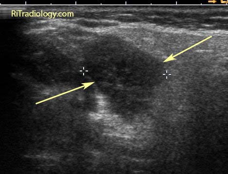
A longitudinal ultrasound image of the kidney shows a focal depression of the lower pole cortex (arrows) with focal parenchymal thinning and a caliceal stone (between calipers).

Facts:
  • Renal scar is a common incidental finding during imaging of the GU tract
  • It can occur both with and without episodes of infundibular obstruction
  • Reflux is considered a major contributor in development of non-obstructive scarring, particularly in children with vesicoureteric reflux (VUR)
  • In adults, renal scarring is more associated with renal stone disease, either with stone or history of stone
Imaging
  • Focal cortical thinning and depression of the cortex, overlying the pyramid on any imaging modalities (IVU, US, CT, MR)
  • Hyperechoic band is seen over the parenchymal thinning on US
  • Mimic = normal renal lobulation. Lobulation will span the pyramids with echogenic lobular junctions into renal columns
Reference:
Newhouse JH, Amis, Jr, ES. The relationship between renal scarring and stone disease. AJR 1988; 151:1153-1156.

November 11, 2011

Parotid Mass


US and CT images of a solid mass in the right parotid gland of an 82-year-old woman who had a painless neck mass for a year.

Facts: Parotid Mass Workup
  • Long list of differential possibilities: neoplastic vs. non-neoplastic conditions
  • Most helpful test = fine needle aspiration biopsy (accuracy 85-90% in experienced cytologist's hands)
  • CT/MRI helpful for treatment planning to determine disease extent and whether facial nerve would need to be sacrificed during surgery
Differential Diagnosis
  • Can be limited by patient's demographic information: age and immune status
  • Facial nerve palsy implies malignancy and poor prognosis
  • Children: hemangioma, lymphangioma, first branchial cleft cyst, pleomorphic adenoma
  • AIDS: benign lymphoepithelial cysts, infection, lymphoma
  • Adults: pleomorphic adenoma (>80%), Warthin tumor, malignant tumor (mucoepidermoid, adenoid cystic carcinoma), metastasis, lymphoma
  • Other nonneoplastic parotid masses: reactive adenopathy, cystic lymphoid hyperplasia, sarcoidosis
Our case: Warthin tumor confirmed by biopsy

References:
1. Steward M, Selesnick SH. Differential Diagnosis in Otolaryngology: Head and Neck Surgery
2. Castillo M. Neuroradiology Companion: methods, guidelines, and imaging fundamentals, 3rd ed, 2006

November 1, 2011

Unilateral Diaphragmatic Elevation

An AP chest radiograph shows elevation of the right hemidiaphragm.

Unilateral Diaphragmatic Elevation: Differentials
  • Lung/pleural disease: Pneumonectomy, lobectomy, pleurisy, subpulmonic effusion
  • Diaphragm disease: Phrenic nerve palsy / eventration
  • Abdominal disease: Hepatomegaly / hepatic mass / abdominal neoplasm / distended stomach


US and CT images demonstrate a very large cyst in the right lobe liver as a cause of elevated right hemidiaphragm.

ShareThis