
 Fig.1 Lateral knee dislocation shown on plain radiograph of a 43-year-old man who fell from height. A bony fragment is noted (red arrow) in the lateral tibiofemoral compartment.
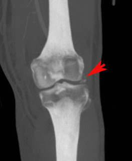
Fig. 2 CT femoral angiography was performed after relocation of knee dislocation to exclude popliteal artery injury. There was no arterial injury. However, CT confirmed a bone fragment in the lateral tibiofemoral compartment of unknown donor site.
Facts:
- Acute knee dislocation (less than 3 weeks), chronic (equal or more than 3 weeks)
- Anterior > posterior > medial, lateral, rotational dislocation
- Current classification scheme uses associated ligamentous injury as a determinant
- Common associated injuries = peroneal nerve injury (20%), popliteal artery injury (19%), fracture of distal femur or proximal tibia (16%)
- When to image popliteal artery? history of ischemia, signs of impaired circulation such as color change, diminished/absent pulses, <>
- Operative management immediate if vascular injury is confirmed. Patterns of ligamentous injury dictate later-stage management.
Reference:
Robertson A, Nutton RW, Keating JF. Dislocation of the knee. J Bone J Surg Br 2006 (June 2006)


No comments:
Post a Comment