
August 30, 2008

August 29, 2008
Gallstone Pancreatitis

 Fig. 1 & 2. Axial and coronal reformatted CT images of a 43-year-old woman who presented with acute epigastric pain and elevated lipase. There is enlarged pancreas with peripancreatic fat stranding (red arrows). Multiple calcified stones are seen in the dilated common bile duct (blue arrows).
- Gallstones = most common cause of acute pancreatitis (Western countries)
- Most stones in common bile duct are from passing of gallstones
- CT can confirm diagnosis of pancreatitis, detect complications of pancreatitis, demonstrate causes of pancreatitis (CBD stone, neoplasm) and exclude other abdominal emergencies
August 26, 2008
Imaging During Pregnancy: What Are the Risks of CT?
 Figure: Sagittal CT image of a pregnant woman who received the scan to rule out acute appendicitis. A gravid uterus (red arrows) with placenta at its fundus, and a fetus were noted. CT was negative for acute appendicitis.
FALSE STATEMENT - "Pregnancy should be terminated after an abdominal CT in early pregnancy."
What really are the risks of doing CT scan in a pregnant woman?
- Spontaneous Abortion: within first 2 weeks of embryonic age (all or none, meaning that failed implantation vs. survival)
- Teratogenesis: 2nd - 20th weeks gestation. Mental retardation, growth restriction, behavioral defect, cataracts. Threshold dose has to be reached to produce an effect. Estimate threshold dose 5-15 rad. Single standard pelvic CT (1-4.6 rad) unlikely to cause teratogenesis.
- Carcinogenesis: always the risk after irradiation of fetus in utero, regardless of dose. Risks higher with exposure in first trimester than with later. Risk increases up to 2 folds at 5 rad of exposure (risk of dying from childhood cancer increases from 1 in 2,000 to 2 in 2,000).
---
การทำ CT ในผู้ป่วยที่ตั้งครรภ์มีความเสี่ยงต่อเด็กในครรภ์ใน 3 รูปแบบด้วยกัน ขึ้นกับอายุครรภ์, ปริมาณของรังสีที่ได้รับ (dose) โดยที่ในช่วง 2 สัปดาห์แรกของการตั้งครรภ์ จะเสี่ยงต่อการแท้ง (ซึ่งถือเป็น all or none phenomenon คือว่าถ้า blastocyst ยังไม่ฝังตัวก็อาจแท้ง แต่ถ้าฝังไปแล้วก็อาจไม่เป็นไร), teratogenesis จะเกิดหรือไม่ขึ้นกับ dose ถ้าถึง threshold (threshold จริงๆ ไม่มีใครทราบแน่ แต่คาดว่าประมาณ 5-15 rad) ในขณะที่ pelvic CT โดยทั่วไป dose ไม่ถึง 5 rad - อันนี้ก็ตรวจสอบกับรังสีแพทย์ที่ทำงานด้วยอีกทีนะครับ เพราะแต่ละสถานที่ก็ใช้เทคนิกแตกต่างกัน ซึ่งอาจทำให้ dose แตกต่างกันด้วย), และ carcinogenesis ซึ่งเกิดได้โดยไม่ขึ้นกับ dose และโอกาสจะสูงกว่าถ้า fetus ได้รับรังสีในช่วงอายุครรภ์น้อยๆ. อย่างไรก็ตาม โอกาสของการเป็นมะเร็งในเด็กที่ทำให้เสียชีวิต โดย baseline ก็ถือว่าน้อยมากอยู่แล้ว (1 ใน 2000) ถ้าได้รับรังสีถึง 5 rad ก็จะเพิ่มความเสี่ยงขึ้น 2 เท่า เป็น 2 ใน 2000 ซึ่งก็ยังถือว่าน้อย.
ต้องแจ้งให้ผู้ป่วยทราบถึงความเสี่ยงและประโยชน์ที่จะได้รับจากการทำ CT scan ก่อนเสมอ
Reference:
1. Chen MM, et al. Obst Gynecol 2008;112(2):333-340 (Aug 2008)
2. McCollough CH, et al. Radiographics 2007;27:909-917. (Jul - Aug 2007).
August 23, 2008
Lateral Knee Dislocation
 Fig.1 Lateral knee dislocation shown on plain radiograph of a 43-year-old man who fell from height. A bony fragment is noted (red arrow) in the lateral tibiofemoral compartment.
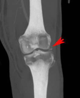
- Acute knee dislocation (less than 3 weeks), chronic (equal or more than 3 weeks)
- Anterior > posterior > medial, lateral, rotational dislocation
- Current classification scheme uses associated ligamentous injury as a determinant
- Common associated injuries = peroneal nerve injury (20%), popliteal artery injury (19%), fracture of distal femur or proximal tibia (16%)
- When to image popliteal artery? history of ischemia, signs of impaired circulation such as color change, diminished/absent pulses, <>
- Operative management immediate if vascular injury is confirmed. Patterns of ligamentous injury dictate later-stage management.
August 20, 2008
August 17, 2008
Radiology Clinical Training in USA (1.4)

- ทางเลือกในการเรียนต่อทางรังสีวิทยา
- ทำไมถึงจะเรียนรังสีวิทยา
- เรียนต่อ Resident เมืองไทย หรือ อเมริกา?
- เรียนต่อ Clinical Fellow, Research Fellow หรือ Observer? เมืองไทย หรือ อเมริกา?
- การเตรียมตัวเพื่อเรียนต่อรังสีวิทยาในสหรัฐอเมริกา
- ตัดสินใจ และเตรียมพร้อม
- USMLE
- การเตรียมตัวสมัครเรียนต่อรังสีิวิทยาในสหรัฐอเมริกา
- การสอบสัมภาษณ์และประกาศผล
- การเตรียมตัวเพื่อเข้าเรียนหลังจากได้รับการตอบรับแล้ว
- ชีวิตการเรียนในอเมริกา
- ชีวิตส่วนตัวในอเมริกา
- แนะนำรุ่นพี่ตัวอย่างที่เป็นแรงบันดาลใจให้กับน้องๆ
- พยายามแล้วไม่สำเร็จ ทำยังไง
Research Fellowship เป็นอีกทางเลือกหนึ่งซึ่งในการมาดูงานและปฏิบัติงานที่อเมริกาครับผู้ที่มาจะทำงานวิจัย (เป็นผู้ช่วยวิจัยครับ แต่ไม่ใช่ ผู้ทำวิจัยหลักเนื่องจากจะไม่สามารถเป็น Principal Investigator ได้) ในงานที่ริเริ่มโดยแพทย์ที่นี่ครับ และในหลายๆ ครั้งก็ได้เข้าร่วม clinical conference ด้วยแล้วแต่ความยุ่งของงานวิจัยและความสนใจส่วนตัวครับ
ข้อดี - มีโอกาสเห็นและร่วมงานวิจัย ซึ่งอาจเป็นงานขนาดใหญ่ และก็ได้เก็บผลงานนี้เวลากลับไปทำงานต่อที่เมืองไทยด้วย (โดยเฉพาะผู้ที่ทำงานในคณะแพทย์ฯ) นอกจากนั้นก็มีโอกาสเข้าร่วม conference ต่างๆ ตามเวลาที่ีมีและความสนใจครับ. ส่วนใหญ่ก็มักเป็นที่โรงพยาบาลหรือมหาวิทยาลัยขนาดใหญ่ครับ ที่เปิดให้มี research fellowship
ข้อเสีย - เวลาที่มีให้กับการเรียนรู้ทางคลินิกจะน้อยกว่าผู้ที่มาเป็น observer เนื่องจากเวลาส่วนใหญ่จะใช้ไปกับการเก็บข้อมูล วิเคราะห์ผลงานวิจัย
Clinical Fellowship
คือการมาปฏิบัติงานเหมือนกับที่รังสีแพทย์ที่อเมริกาทำงานครับ และได้รับเงินเดือนจากโรงพยาบาลในอเมริกาที่เราทำงานครับ เช่น การอ่านผล imaging การออกตรวจผู้ป่วย (กรณีทำ ultrasound, fluoroscopy หรือ intervention) การพูดคุยกับผู้ป่วย เป็นต้น นอกจากนั้นก็มีโอกาสทำงานวิจัย (ถ้าสนใจ)
ข้อดี - มีโอกาสสัมผัสงาน และได้ปฏิบัติงานจริงเหมือนกับเป็นรังสีแพทย์ครับ ทำให้มีการเรียนรู้โดยตรงครับ ได้ประสบการณ์ตรงทั้งในแง่ของการทำงานและระบบของโรงพยาบาลที่เราทำงาน ได้รู้จักและทำงานกับรังสีแพทย์ที่หลากหลาย ทั้ง resident, fellow และ attending มีโอกาสทำงานวิจัยและเป็น principal investigator
ข้อเสีย - โรงพยาบาลขนาดใหญ่ งานก็ยุ่งตามจำนวนผู้ป่วยครับ หลายๆแห่งต้องอยู่เวรวันหยุด หรือข้ามคืน (ขึ้นกับโปรแกรม). ต้องสอบ USMLE ให้ผ่านทั้งหมด และผ่านกระบวนการแบบเดียวกับ US resident เวลาสมัครเข้าเรียนต่อครับ. บางโปรแกรมอาจต้องสอบข้อสอบของ fellow ด้วยก่อนจะเรียนจบครับ
ถ้าถามความเห็นส่วนตัวและต้องให้คำแนะนำกับผู้ที่กำลังวางแผนจะมา ก็คงต้องบอกว่าผมแนะนำให้มาเป็น Clinical Fellow> Research Fellow> Observer ครับ แต่ทั้งนี้การเลือกมาขึ้นกับหลายปัจจัยนะครับ ทั้งในแง่ของเวลา เงินทอง ครอบครัวและความตั้งใจ ครับ
บทความต่อๆ ไปจะกล่าวถึงกรณีที่สนใจจะมาเป็น Clinical Fellow หรือ Resident นะครับ
บทความชุดนี้ ผมขอมอบให้กับคนหลายคนที่เป็นแรงบันดาลใจ ให้ผมมาได้จนถึงจุดที่อยู่ปัจจุบันครับ พ่อแม่ น้อง ที่สนับสนุนทุกทางไม่ว่าจะเป็นเรื่องเงินทอง เวลา และกำลังใจ. อ.จิรพร เหล่าธรรมทัศน์ และ อ.จามรี เชื้อเพชรโสภณ รุ่นพี่ตัวอย่างที่ประสบความสำเร็จอย่างสูง ทั้งทางวิชาการและการบริหารจัดการ. อ.วิทย์ วราวิทย์, อ.ชีวรัตน์ วิโรจน์ธนุกูล, พี่นัศวดี เพียงเจษฎา, บัณฑิต ตันติวงโกสีย์ ที่เป็นผู้ช่วยเหลือตั้งแต่ตอนเริ่มคิด สอบ และส่งแรงให้ผมได้มาอยู่ตรงนี้
August 14, 2008

August 11, 2008
Three 'Reasonable and Appropriate' Indications for Coronary CTA for Detection of CAD
 กล่าวตามบรรยายของ Dr. Mamuya ซึ่งเป็น MGH cardiologist รับเชิญมาพูดเกี่ยวกับ Clinical Role of Cardiac CTA ที่เพิ่งผ่านมาเร็วๆ นี้
Indications ที่เหมาะสมในการทำ Coronary CTA for detection of CAD มี 3 ข้อ
- Symptomatic patients with intermediate pretest probability who have either uninterpretable EKG or unremarkable EKG
- Symptomatic patients with uninterpretable stress test
- Symptomatic patients with new onset of heart failure
สรุปว่า
Coronary CTA is appropriate in SYMPTOMATIC patients with INTERMEDIATE pretest probability
Reference:
Hendel, et al. ACCF/ACR/SCCT/SCMR/ASNC/NASCI/SCAI/SIR Appropriateness Criteria for Cardiac Computed Tomography and Cardiac Magnetic Resonance Imaging. JACC (2006)
August 8, 2008
How Doctors Think: Chapter 3
 "In order to think well, especially in hectic circumstances, you need to slow things down to avoid making cognitive errors."
บทที่ 3 ของหนังสือเล่มนี้เล่าเรื่องของหมอ ER ครับ เริ่มด้วยเรื่องของ Nathan ผู้ป่วยเด็กอายุ 10 ปีซึ่งมาตรวจที่ ER ของ โรงพยาบาลใน Nova Scotia ด้วยอาการปวดหลังฉับพลัน เนื่องจากมี compression fracture ของ T10 หมอผู้ตรวจเกิดความสงสัยว่าอาจจะเป็น pathologic fracture จึงได้ส่งตรวจ CT และ MRI ต่อ ก็พบว่าความผิดปกติมีอยู่ที่ T10 ที่เดียว จึงโทรไปปรึกษาหมอเด็ก และได้รับคำตอบว่าจากหมอเด็กว่าสามารถพบได้. ด้วยเหตุที่ 'specialist' ว่ามา จึงได้ให้ Nathan กลับบ้านไป. 2-3 สัปดาห์ถัดมา Nathan ก็มาที่ ER อีกครั้งด้วยอาการเดิม เอกซเรย์พบว่ามี compression fractures ในตำแหน่งใหม่เพิ่มขึ้นอีก คราวนี้หมอ ER ส่ง Nathan ไป biopsy พบว่า Nathan ป่วยเป็น Acute Lymphoblastic Leukemia (ALL)
เรื่องของ Nathan สอนให้รู้ว่า แม้ว่าจะเป็น specialist ก็พลาดได้ด้วยประโยคสั้นๆ ง่ายๆ ว่า "We see this sometimes." (ของแบบนี้พบได้ - ไม่แปลก). จริงๆ แล้วก่อนที่เรา (ในฐานะที่เป็นหมอ) จะพูดประโยคดังกล่าว ต้องคิดให้รอบคอบอยู่เสมอว่าเราได้พยายามจนถึงที่สุดในการค้นหาการวินิจฉัยแล้ว ถ้ายังไม่ถึงที่สุด ก็ไม่ควรใช้ความคิดดังกล่าวมายุติการค้นหาคำตอบ
ในบทนี้ Dr. Groopman ยังได้เล่าเรื่องราวผ่านคนไข้หลายๆ คน เช่น Blanche ซึ่งมาตรวจที่ ER ด้วยอาการหอบเหนื่อย มีไข้ ในช่วงเวลาที่ชุมชนใกล้เคียงมีการระบาดของไวรัส ทั้งๆ ที่ lab กับ x-ray ของ Blanche ดูปกติแต่หมอ ER ก็วินิจฉัยว่าเป็น Viral Pneumonia. ปรากฎว่าที่แท้จริงแล้ว Blanche ป่วยเนื่องจาก Aspirin Toxicity. อีกเรื่องที่น่่าสนใจเป็นคนไข้ที่ป่วยด้วย Irritable Bowel Syndrome มาด้วยอาการปวดท้องฉับพลันที่ ER ถึง 3 ครั้ง และถูก "ตราหน้า" ว่าเป็น Functional diagnosis. ปรากฎว่าผู้ป่วยถูกพามายัง ER เป็นครั้งที่ 4 ด้วยอาการ shock และพบว่าเป็น Ruptured Ectopic Pregnancy. ผู้เขียนได้ชี้ให้เห็นว่า Confirmation bias มีบทบาทสำคัญทำให้หมอวินิจฉัยโรคผิดพลาด เนื่องจากหมอเลือกที่จะดูข้อมูลบางอย่าง และ เลือกที่จะไม่สนใจข้อมูลบางอย่างของผู้ป่วย
หลายๆ จุดในหนังสือเล่มนี้ ทำให้ผมมองเห็นว่าในฐานะที่เรา (ส่วนมากของผู้อ่าน) เป็น radiologistและเป็น diagnostician เช่นกัน น่าจะได้ประโยชน์ในการเปิดมุมมองว่า ทำไมในหลายๆ ครั้ง เราจำเป็นต้อง definite แต่บางครั้งก็ต้องให้ differential diagnosis จากข้อมูลที่มีในภาพ imaging เนื่องจากเราก็มี bias ได้ไม่ต่างจาก clinicians
----
In chapter 3, Groopman described lives of emergency physicians and errors in diagnosis due to bias. Nathan, a 10-year-old boy arrived at the ER of Nova Scotia hospital with an acute back pain. An x-ray showed a T10 compression fracture. ER doctor consulted a pediatrician and received a message "We see this sometimes". He let Nathan go home to be found out later that Nathan had an acute leukemia. It is important, as emphasized by Groopman, that any doctors (even specialists) should be certain of their words before they dismiss any findings of the patients. He also gave examples of confirmation bias through a patient who was diagnosed as viral pneumonia (she actually had aspirin toxicity) in the period of influenza epidemic, and another IBS patient who was repeatedly diagnosed as functional abdominal pain (she actually had ruptured ectopic pregnancy).
Jerome Groopman ปัจจุบันเป็น Professor (medicine) ที่ Beth Israel Deaconess Hospital (Boston). เขาจบแพทย์จาก Harvard Medical School และทำ internship กับ medicine ในรั้ว Harvard ที่ Massachusetts General Hospital. เขาเคยทำงานที่ UCLA Medical Center ก่อนจะย้ายกลับมาอยู่ที่บอสตัน.
August 5, 2008
Isolated Ulnar Styloid Process Fracture
 รูปบน - Oblique radiograph of the wrist shows a fracture of the tip of the ulnar styloid process. There is no associated radial fracture.
Ulnar styloid process fracture ส่วนมากมักพบร่วมกับ distal radius fracture แต่บางครั้งก็พบเป็น isolated fracture ได้ดังเช่นในเคสตัวอย่าง. Ulnar styloid fracture มี 2 แบบ แบ่งตามตำแหน่งของ fracture ว่าอยู่ที่ base หรือ tip ของ styloid process.
ข้อควรรู้
- Fracture ที่ base พบว่า associated กับ instability of distal radioulnar joint (DRUJ), Triangular fibrocartilage (TFCC) injury และ rate of nonunion สูงกว่า fracture ที่ tip
Hauck RM, et al. Classification and treatment of ulnar styloid nonunion. J Hand Surg [Am] 1996;21:418-422.
August 2, 2008
Appendiceal Mucocele

 Fig. 1 and 2: Axial (1) and sagittal (2) CT images of a 55-year-old man with right lower quadrant pain show a sausage-shaped cystic lesion (red arrows) with a thin rim of calcification (blue arrow) in the appendix.
- Mucocele is an abnormal accumulation of mucus in a dilated appendix.
- Three distinct pathology of mucocele are 1. mucosal hyperplasia, 2. mucinous cystadenoma (most common) and 3. mucinous cystadenocarcinoma. They are indistinguishable on imaging.
- Preoperative diagnosis is important. Careful handling during surgery reduces a risk of rupture and development of pseudomyxoma peritonei.
- CT findings = low-attenuation mass, smooth thin or thick wall.
Mucocele ของ appendix สันนิษฐานว่าเกิดจากการอุดตันของ appendix ทำให้มี mucus สะสมอยู่ภายใน ก่อให้เกิดอาการเช่น ก้อน ปวดท้องน้อยด้านขวา เป็นต้น. Mucocele อาจเป็นได้ตั้งแต่ benign ไปจนถึง malignant ซึ่งอาจแยกไม่ได้ด้วย imaging ดังนั้นการผ่าตัดจึงมีบทบาททั้งในแง่การวินิจฉัยและการรักษา. การที่ surgeon รู้ว่าเป็น mucocele มีประโยชน์ในแง่ที่ช่วยเพ่ิมความระมัดระวังระหว่างการผ่าตัด เพราะถ้าแตกเข้าไปในช่องท้อง ผู้ป่วยจะเสี่ยงต่อการเกิด pseudomyxoma peritonei.
Reference:
Kim SH, et al. Mucocele of the appendix: ultrasonography and CT findings. Abdom Imaging 1998; 23:292-296.


