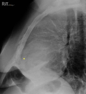
 Figure 1: Lateral chest radiograph shows separation of the retrosternal (dark line parallel to the sternum anterior to the yellow star) and epicardial fat stripes (dark line behind the yellow star). This patient also has an anterior mediastinal mass due to lymphoma.
 Figure 2: Axial contrast-enhanced CT image shows a large pericardial effusion (stars) separating the retrosternal fat stripe (double-headed arrow) and epicardial fat stripe (arrowheads).
Facts: Pericardium
- Pericardium has two layers: visceral (attached to myocardial surface and proximal great vessels) and parietal (free wall of pericardial sac)
- Pericardial sac normally contains 20-50 mL of fluid
Facts: Pericardial Effusion
- Most common cause = myocardial infarction with left heart failure
- Other causes: uremia, hypoalbuminemia, myxedema, infection, drug reaction, trauma, neoplasm, autoimmune disease
- Can be seen on radiography if volume exceeds 250 mL
Imaging Features
- PA or AP radiograph: water bottle-shaped morphology of the cardiomediastinal shadow
- Lateral view: separation of retrosternal and epicardial fat stripes by more than 2 mm (Oreo cookie sign)
- Oreo cookie sign: epicardial fat and retrosternal fat stripes = outer dark cookie layers; opaque fluid = white fluff of the cookie
Reference:
Parker MS, Chasen MH, Paul N. Radiologic signs in thoracic imaging: case-based review and self-assessment module. AJR 2009;192:S34-S48.
Follow RiTradiology on Facebook, Twitter or Google Friend Connect
Visit RiT Illuminations to view nice pictures of your colleague


No comments:
Post a Comment