December 21, 2012

Atypical Hepatic Hemangioma - Rapid Filling of Contrast

Axial MR images in multiple phases (as labeled) show a T1 hypointense nodule in the right hepatic lobe that rapidly filled in with contrast after administration and subsequently fades on delayed imaging. Note that the enhancement of the nodule is similar to the aorta in all phases. On T2W image (not shown), this nodule has a very high signal intensity. 


Rapidly Filling Hemangioma
  • 16% of all hepatic hemangiomas
  • More common among small (<1 1="1" 42="42" cm="cm" hemangioma="hemangioma" hemangiomas="hemangiomas" in="in" incidence="incidence" less="less" li="li" of="of" than="than">
  • Immediate homogeneous enhancement at arterial phase and hyperintensity persists in delayed phases. Enhancement equal to aorta in all phases. 
  • High T2 signal intensity may be helpful for differentiation from other arterial enhancing nodules (but it can also be seen in islet cell tumor metastasis)

Reference:
Vilgrain V et al. Imaging of atypical hemangiomas of the liver with pathologic correlation. Radiographics 2000; 20:379

December 11, 2012

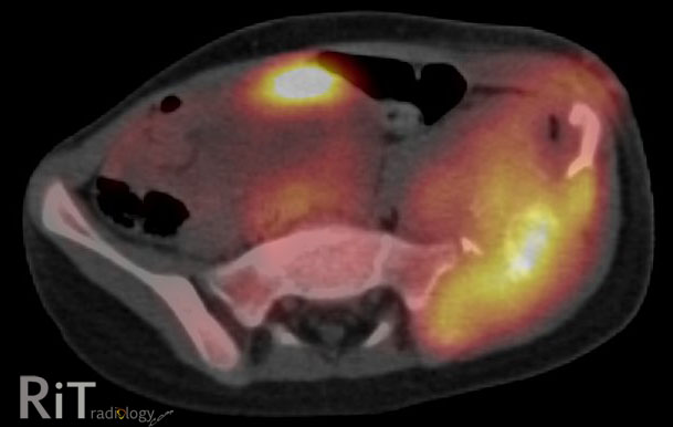
Pelvic Ewing's Sarcoma

A pelvic radiograph of a 5-year-old girl shows a large lytic lesion in the left iliac bone (arrows).
An axial FDG PET/CT image shows high metabolic activity of the mass involving the left iliac bone with soft tissue component and bone destruction. A coronal T2W MR image reveals an extensive soft tissue mass with necrotic areas and involvement of the adjacent musculature. 


Differential Diagnosis
  • Metastatic neuroblastoma. Given her age at five years old, this needs to be in differentials
  • Ewing's sarcoma
  • Telangiectatic osteosarcoma
  • Osteomyelitis. Great mimics of aggressive-looking bone tumor. Symptoms may overlap with round-cell tumor, including fever
This case: Ewing's sarcoma by tissue diagnosis (+ve PAS and vimentin).

Facts: Ewing's Sarcoma

  • Malignant round-cell tumors of the bone with neural cell origin
  • Tumors of children and young adults, most between 10-20 years old. Less than 2% occur in children less than 5 years old
  • Most common sites = femur >> pelvis
  • Pelvic Ewing's -- bad prognosis because there is no anatomic barrier to tumor spread, close proximity to viscera and neurovascular bundles, prone to recur

Reference:
Bhagat S, Sharma H, Pillai DS, Jane MJ. Pelvic Ewing's sarcoma: a review from Swedish Bone Tumour Registry. J Orthop Surg 2008;16:333-8

November 30, 2012

Maisonneuve Fracture

Figure 1: AP and lateral ankle radiographs demonstrate a vertical fracture of the medial/posterior malleolus of the distal tibia without a fibular fracture.  

Figure 2: Full-length AP fibular radiograph shows a mildly displaced fracture of the fibular shaft at the junction between the proximal 1/3 and middle 1/3. 

Facts: Rotational Ankle Fractures

  • Rotational ankle fractures are classified according to force direction applied to the foot, while the injured foot can be in a different position (supination/pronation, adduction/external rotation)
  • AO/Weber classification: A, B, C fractures are differentiated by location of fibular fractures. 
  • Fibular fracture below the syndesmosis = AO/Weber A (usually supination-adduction)
  • Fibular fracture at the syndesmosis = AO/Weber B (~ supination and external rotation)
  • Fibular fracture above the syndesmosis = AO/Weber C (~ pronation external rotation)
Facts: Maisonneuve Fractures
  • High fibular fracture above the syndesmosis resulting from external rotation
  • Often, there is injury to the medial ankle either a tranverse medial malleolar fracture, posterior malleolar fracture or disruption of the deltoid ligament
  • Disruption of the syndesmosis and interosseous ligament up to the fibular fracture site
  • Suspicious for this fracture if you see a 1) transverse medial malleolar fracture or 2) posterior malleolar fracture but no fibular fracture on the ankle radiographic series. In these situation, a full-length fibular radiograph should be taken

Reference:
Sakthivel-Wainford K. Self-assessment in limb x-ray interpretation, 2006
Rockwood CA, Green DP. Rockwood and Green's fractures in adults, 2005


November 26, 2012

Screening Mammography Overdiagnoses Breast Cancer?

The recently published NEJM's original article describes effect of screening mammography on breast cancer incidence in the USA. Bleyer A and Welch HG. N Eng J Med 2012;367:1998 (22 Nov 2012)


Facts: Aim of Cancer Screening

  • We expect cancer screening program to lower cancer-related mortality by a means of 1) earlier detection of disease destined to be fatal and 2) early Rx of screen-detected cancers
  • A cancer screening program should 1) increase incidence of cancer detected at an earlier stage and 2) decrease incidence of cancer presenting at a late stage

What Study Is About And What It Has Found:
  • Breast cancer rate from 1976 through 1978 (mammography was uncommon) ~ baseline incidence
  • Breast cancer rate from 2006 through 2008 ~ current incidence
  • Confounding effects of menopausal hormone therapy were minimized by not including transitory increase in incident breast cancers from 1990 through 2005. Underlying incidence of breast cancer assumed to rise by 0.25% annually
  • Number of cases of early-staged breast cancer rises from 112 to 234 cases per 100,000 women
  • Number of cases of late-staged breast cancer decreases from 102 to 94 cases per 100,000 women
  • "With an assumption of a constant underlying disease burden, only 8 of the 122 additional early-stage cancers diagnosed were expected to progress to advanced disease." This was estimated that breast cancer was overdiagnosed. In 2008, the overdiagnosis would account for 31% of all breast cancer diagnosed
Other Studies Said?
Results of this study concur with other reports (references 2-3) and suggest that improvements in treatment are the main drive in reducing breast cancer mortality - rather than screening mammography. 

It is suggested that "pros and cons of mammography should be incorporated into the counseling that women receive as they decide whether an when to be screened". 


References:
  1. Bleyer A and Welch HG. Effect of three decades of screening mammography on breast cancer incidence. N Eng J Med 2012;367:1998
  2. Kalager M et al. Effect of screening mammography on breast cancer mortality in Norway. N Eng J Med 2010;363:1203
  3. Autier P et al. Breast cancer mortality in neighbouring European countries with different levels of screening but similar access to treatment: trend analysis of WHO mortality database. BMJ 2011;343:d4411
  4. Kaunitz AM. Screening mammography: does overdiagnosis overshadow prevention of advanced breast cancer? Journal Watch Women's Health November 21, 2012

November 20, 2012

Spinal Langerhans Cell Histiocytosis (LCH)

Figure 1: Tc-99m MDP bone scan (posterior image) shows a focus of increased activity at L2 vertebral body in a 24-year-old woman presenting with back pain.

Figures 2&3: Axial GRE T2W and sagittal post-contrast T1W MR images show a round focus of bone destruction surrounded by bone marrow edema and enhancement of L2 body, sparing the posterior elements. 

Facts: LCH
  • Rare, benign disorder of unknown etiology comprises of eosinophilic granuloma (unifocal), Hand-Schuller-Christian disease (multifocal) and Letterer-Siwe disease (disseminated variant) - these are different manifestations of a same disease
  • Clonal proliferation of Langerhans cells
  • Peak incidence 5-10 years but there is a shift toward younger children
Spinal LCH
  • Vertebral involvement in 8-25% of cases
  • Thoracic > cervical, lumbar
  • Vertebral body >> posterior elements
  • Solitary, well-defined osteolytic lesion with scalloped borders eventually progresses to collapse and a classic "vertebra plana"
  • Typically single vertebral body involved. Disc spaces spared
  • Soft tissue mass suggests more aggressive course
Main Imaging Differentials of Spinal LCH
  • Osteomyelitis
  • Ewing sarcoma
  • Leukemia, lymphoma, metastatic neuroblastoma

Reference:
Hosalkar HS, Greenberg JS, Wells L, Dormans JP. Isolated Langerhans Cell Histiocytosis of the T12 vertebra in an adolescent. Am J Orthop 2007;36: E21-E24.

ShareThis