December 21, 2011

Sternoclavicular Rheumatoid Arthritis


Axial CT images of the sternoclavicular joints show erosion, indistinct cortical margins of the joints, which are quite symmetric.

Facts:
  • Sternoclavicular (SC) joint is diarthrodial articulation between axial and appendicular skeleton, which is subject to same disease process that occur in other joints (degenerative arthritis, rheumatoid arthritis, infection and subluxation. Degenerative arthritis is the most common).
  • Up to 30% of patients with rheumatoid arthritis have changes in SC joints (a part of polyarticular involvement) but radiographic findings are often unremarkable.
Imaging Finding
  • Synovitis
  • Bone marrow edema and enhancement of subcortical bone (after IV contrast)Er
  • Erosion and indistinct cortical margins
  • Think infection if: unilateral, history of IVDU and immunocompromised states
References:
1. Restrepo CS, Martinez S, Lemos DF, et al. Imaging appearance of the sternum and sternoclavicular joints. Radiographics 2009; 29:839-859.
2. Berry DJ, Steinmann SP. Adult Reconstruction, 2007.

December 11, 2011

Scrotal Pyocele

Gray-scale and color Doppler US images of the testicle shows a complex fluid collection (stars) around the testicle and marked scrotal skin thickening. The epididymis is edematous with increased flow (image not shown).

Facts: Scrotal Pyocele
  • Also known as scrotal abscess
  • Can be superficial (from infected hair follicles, wound) or intrascrotal
  • Causes: epididymitis, TB, instrumentation, neurogenic bladder, chronic catheter indwelling, spread from intraabdominal infection (i.e., appendicitis)
  • Intrascrotal abscess requires surgical drainage
Imaging
  • US is the modality of choice
  • Complex-appearing fluid around the testicle
  • Scrotal skin thickening with hyperemia
  • Evidence of causes such as epididymitis or others
  • Based on imaging, it is difficult to distinguish pyocele from hematocele
References
1. Siegel MJ. Pediatric Sonography, 2010.
2. Resnick MI, Novick AC. Urology Secrets, 3rd ed, 2003.

November 21, 2011

Renal Scarring

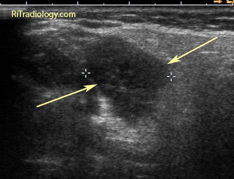
A longitudinal ultrasound image of the kidney shows a focal depression of the lower pole cortex (arrows) with focal parenchymal thinning and a caliceal stone (between calipers).

Facts:
  • Renal scar is a common incidental finding during imaging of the GU tract
  • It can occur both with and without episodes of infundibular obstruction
  • Reflux is considered a major contributor in development of non-obstructive scarring, particularly in children with vesicoureteric reflux (VUR)
  • In adults, renal scarring is more associated with renal stone disease, either with stone or history of stone
Imaging
  • Focal cortical thinning and depression of the cortex, overlying the pyramid on any imaging modalities (IVU, US, CT, MR)
  • Hyperechoic band is seen over the parenchymal thinning on US
  • Mimic = normal renal lobulation. Lobulation will span the pyramids with echogenic lobular junctions into renal columns
Reference:
Newhouse JH, Amis, Jr, ES. The relationship between renal scarring and stone disease. AJR 1988; 151:1153-1156.

November 11, 2011

Parotid Mass


US and CT images of a solid mass in the right parotid gland of an 82-year-old woman who had a painless neck mass for a year.

Facts: Parotid Mass Workup
  • Long list of differential possibilities: neoplastic vs. non-neoplastic conditions
  • Most helpful test = fine needle aspiration biopsy (accuracy 85-90% in experienced cytologist's hands)
  • CT/MRI helpful for treatment planning to determine disease extent and whether facial nerve would need to be sacrificed during surgery
Differential Diagnosis
  • Can be limited by patient's demographic information: age and immune status
  • Facial nerve palsy implies malignancy and poor prognosis
  • Children: hemangioma, lymphangioma, first branchial cleft cyst, pleomorphic adenoma
  • AIDS: benign lymphoepithelial cysts, infection, lymphoma
  • Adults: pleomorphic adenoma (>80%), Warthin tumor, malignant tumor (mucoepidermoid, adenoid cystic carcinoma), metastasis, lymphoma
  • Other nonneoplastic parotid masses: reactive adenopathy, cystic lymphoid hyperplasia, sarcoidosis
Our case: Warthin tumor confirmed by biopsy

References:
1. Steward M, Selesnick SH. Differential Diagnosis in Otolaryngology: Head and Neck Surgery
2. Castillo M. Neuroradiology Companion: methods, guidelines, and imaging fundamentals, 3rd ed, 2006

November 1, 2011

Unilateral Diaphragmatic Elevation

An AP chest radiograph shows elevation of the right hemidiaphragm.

Unilateral Diaphragmatic Elevation: Differentials
  • Lung/pleural disease: Pneumonectomy, lobectomy, pleurisy, subpulmonic effusion
  • Diaphragm disease: Phrenic nerve palsy / eventration
  • Abdominal disease: Hepatomegaly / hepatic mass / abdominal neoplasm / distended stomach


US and CT images demonstrate a very large cyst in the right lobe liver as a cause of elevated right hemidiaphragm.

ShareThis