July 30, 2009

Enchondroma of the Foot

Figure 1: AP radiograph of the left foot shows an incidental geographic lytic lesion without sclerotic border at the proximal phalanx of the third toe. There is cortical thinning without breakthrough or associated soft tissue mass. No matrix is visualized.
Figure 2: Sagittal MR image (STIR sequence) shows the lesion as markedly T2 hyperintense without associated soft tissue mass.


Facts
  • Enchondroma is common (10% of all benign bone tumors)
  • Most common in hands and feet
  • Occurs from cartilaginous cell nests displaced from physis during development
  • Often asymptomatic and found incidentally or after a pathologic fracture
  • Main differential diagnosis is chondrosarcoma, which is relatively rare in the foot - and typically arises de novo. However, differentiation using clinical and radiographic parameters can be difficult.
  • Presence of soft tissue mass, cortical destruction and deep endosteal scalloping - although suggests chondrosarcoma - can be seen in both benign and malignant cartilage tumors of the foot
When to Suspect Chondrosarcoma Arising from Enchondroma of the Foot?
  • Lesions larger than 5 cm
  • Midfoot or hindfoot
  • These lesions should be considered malignant and biopsy or close clinical follow up recommended

Reference:
Gajewski DA, Burnett JB, Murphy MD, Temple HT. Differentiating clinical and radiographic features of enchondroma and secondary chondrosarcoma in the foot. Foot Ankle Int 2006;27:240-242.

July 27, 2009

Fibromatosis Colli of Infancy

Transverse ultrasound images of the right sternocleidomastoic (SCM) muscle and left (symptomatic side) shows diffuse enlargement of the left SCM with mixed echogenicity in a 2-month-old infant.


Facts: Fibromatosis Colli of Infancy
  • Abnormal mass-like enlargement of sternocleidomastoic (SCM) muscle
  • Born normal but develops SCM mass and/or torticollis within the first 4-8 weeks of life
  • Associated with difficult delivery and intrauterine torticollis due to malposition
  • Histology: collagen fibers and fibroblasts around atrophied muscle fibers
  • Early diagnosis and treatment important because it can result in contracture
  • Responds well to physiotherapy
Ultrasound Appearance
  • US performed to exclude other diagnosis such as cervical lymphadenopathy, rhabdomyosarcoma, lymphoma, cystic hygroma or branchial cleft cyst
  • Mass or fusiform enlargement of the SCM muscle
  • Confined to muscle, no adjacent abnormalities of other soft tissues
  • Variable echogenicity -- hyperechoic, hypoechoic, or mixed echogenicity
Reference:
Bedi DG, John SD, Swischuk LE. Fibromatosis colli of infancy: variability of sonographic appearance. J Clin Ultrasound 1998;26:345-348.

July 24, 2009

Embryonic Demise

Figure 1: Transvaginal ultrasound shows a mean sac diameter of 30 mm (about 8 weeks gestational age). S = gestational sac.
Figure 2: Further scan demonstrates a fetal pole (arrow) without a yolk sac. There is minimal decidual reaction.


Embryonic Demise
  • Most common cause = chromosomal abnormality leading to arrested development
  • Diagnosis is made by:
  1. CRL > 5 mm but no cardiac activity
  2. MSD >/= 8 mm but no yolk sac
  3. MSD >/= 16 mm but no embryo
  4. Embryo seen but no yolk sac
  5. No cardiac activity in embryo seen by transabdominal ultrasound (need to confirm with transvaginal ultrasound if early IUP)

Reference:
Weissleder, Wittenberg, Harisinghani, Chen. Primer of diagnostic imaging. 4th edition, 2007

July 21, 2009

Unilateral Opacity of a Hemithorax

Figure 1: Supine chest radiograph shows a unilateral increased opacity in the left hemithorax, particularly at the mid to lower zones. There is no mediastinal shift. The left hemidiaphragm is in a normal position.


Differential Diagnosis
  1. Lung: consolidation, mass, collapse
  2. Pleura: effusion (layering or loculated), mass (metastasis, mesothelioma)
  3. Chest wall: mass (breast, chest wall musculatures)
  4. Extrathoracic: external structures

In this case, the opacity extends beyond the lung to the soft tissue of the chest wall, corresponding to the breast. It is unlikely to be a lung lesion because normal pulmonary vasculatures are visualized through it. Pleural lesion would not extend beyond the thoracic cage. Two main possibilities include a large breast mass or external structures (for example, nonradiopaque pads on the bed).

Comparison with the previous chest radiographs revealed that this is a new finding. Further query of patient's history confirmed the diagnosis of a large left breast mass. Chest CT scan (below) also shows the mass (invasive cancer).


Reference:
Chapman S and Nakielny R. Aids to radiological differential diagnosis. 4th edition, 2003.

July 18, 2009

Double Bubble Sign

Supine abdominal radiograph of a newborn presenting with billous vomiting shows a distended stomach and duodenal air bubbles "double bubble" sign. There is no gas distal to the duodenal bubble. Radiopaque materials overlying the right pelvis are due to soiled diaper.


Double Bubble Sign
  • Radiographic signs describing two air-filled structures in the upper abdomen with little or no air distally
  • Can be reproduced in upper GI study or ultrasound
  • Left-sided bubble = stomach
  • Right-sided bubble (usually right of midline) = duodenum
  • Most common cause = duodenal atresia
Differential Diagnoses
  • Intrinsic cause: duodenal atresia, duodenal stenosis, duodenal web
  • Extrinsic cause: annular pancreas, malrotation with obstruction produced by midgut volvulus or by Ladd bands
Main Concern
  • Must be differentiated from 'malrotation with obstruction (midgut volvulus)' because this condition requires urgent surgery
  • Suspect midgut volvulus if stomach is distended but duodenum is normal or only slightly dilated

Reference:
Traubici J. The double bubble sign. Radiology 2001;220:463-464.

July 15, 2009

Lipomatous Hypertrophy of Interatrial Septum

Figure: Axial chest CT image (non cardiac-gating) shows a fatty mass (arrows) in the interatrial septum with a "dumbbell shape" sparing the fossa ovalis (double-headed arrow). The mass extends to the wall of the superior vena cava.


Facts
  • Excessive deposition of fat in the interatrial septum of unknown etiology
  • Not common, incidence about 1% in autopsy, 2% on CT and up to 8% on echocardiography
  • In most cases, they are incidental. However, they may cause arrhythmias

CT Appearance
  • Fatty mass with thickness >/= 20 mm, sharp margin, no enhancement
  • Spares fossa ovalis, resulting in a "dumbbell" shape on axial images
  • Fat can extend to the level of coronary sinus and to the aortic root

Imaging Differential Diagnoses
  • Atrial myxoma: this tumor arises from fossa ovalis and is usually pedunculated
  • Cardiac lipoma: encapsulated, true neoplasm in younger patients

Reference:
Heyer CM, Kagel T, Lemburg SP, Bauer TT, Nicolas V. Lipomatous hypertrophy of the interatrial septum: A prospective study of incidence, imaging findings, and clinical symptoms. Chest 2003; 124:2068-2073.

July 12, 2009

Seronegative Spondyloarthropathy (SNSA)

Figure 1: AP radiograph of the lumbosacral spine shows symmetric sclerosis and fusion (ankylosis) of the sacroiliac joints (red arrowheads). There is ossification of the outer fibers annulus fibrosus (arrows).
Figure 2: Lateral view of the spine of the same patient demonstrates ossification of the outer fibers of annulus fibrosus (arrows), squaring of anterior vertebral margin, and "shiny corner" sign (arrowhead).


Facts
  • SNSA is a group of diseases that general pathology consists of negative rheumatoid factor (RF) and elevated erythrocyte sedimentation rate (ESR)
  • Ankylosing spondylitis, reactive spondyloarthropathy, psoriatic spondyloarthropathy, spondyloarthropathy associated with inflammatory bowel disease, undifferentiated spondyloarthropathy
  • Strong association with HLA-B27 haplotype
  • Most common clinical presentation is low back pain (greater in the morning) and pathologic fracture
Imaging Findings
  • First involves sacroiliac joints (erosion of synovial portion, then fusion in late stage), then thoracolumar junction and may involve whole spine
  • Corner erosions of vertebral bodies produce "squaring" and reactive sclerosis produces "shiny corner" sign
  • "Bamboo spine" caused by thin ossification of the outer fibers of annulus fibrosus at vertebral margin, common in ankylosing spondylitis

Key Differentiating Imaging Features
  • Involvement of SI joints - rare in rheumatoid arthritis, normal in DISH, retinoid therapy
  • Infection of SI joint typically unilateral
Our case: ankylosing spondylitis

Reference:
Ross, Brant-Zawadzki, Moore, et al. Diagnostic Imaging: Spine, 2004

July 9, 2009

Rounded Atelectasis

Fig. 1: Lateral chest radiograph of a 77-year-old man, status post recent CABG, shows a round subpleural opacity (red star) in the posterior aspect of the lower lobe. There are small bilateral pleural effusions, left (yellow star) greater than right.
Fig. 2: Axial CT image of the same patient shows a subpleural "masslike" opacity (red star) in the posterior left lower lobe with curvilinear opacities connecting the inner part of the "mass" to the pulmonary hilum (arrows). There is associated volume loss of the left lower lobe and associated left pleural effusion (yellow stars).


Facts:
  • Rounded atelectasis is an unusual form of lung collapse that can simulate lung neoplasm on chest radiography
  • Commonly occurs in patients with pleural effusions/thickening
  • Mostly attributed to asbestos exposure, but other causes have been identified (i.e. TB, infarction, trauma, uremia, postoperative)
  • Men more common than women, average age 60 years
  • Pathology = thickened visceral pleura with folding of underlying lung parenchyma. No discrete mass
  • FNA is usually unnecessary given characteristic imaging findings
Radiologic Findings
  • Rounded, oval (less commonly irregular) subpleural mass
  • Curvilinear opacities connecting the mass to pulmonary hilum (Comet-tail sign)
  • Volume loss of affected lobe (most common in posterior lower lobe)
  • Associated pleural thickening
Reference:

Batra P, Brown K, Hayashi K, Mori M. Rounded atelectasis. J Thorac Imaging 1996;11:187-197.

July 6, 2009

Choledochal Cyst


Fig.1: Transverse ultrasound image at the region of the gallbladder (arrow) shows a nearby large cystic structure (star).
Fig. 2 and 3: Axial T2 MR image and MRCP confirms the presence of the large cystic lesion adjacent to the gallbladder (arrow), proven to be saccular dilatation of the extrahepatic bile duct.

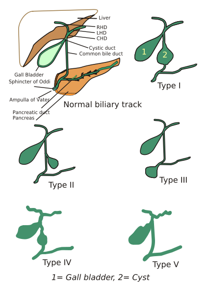
Fig.4: Diagram showing Todani classification of choledochal cyst

Choledochal Cyst
  • Rare, congenital dilatation of biliary tree
  • Can be either intra-hepatic, extra-hepatic or both
  • Female:male ratio = 4:1, more common in Asia
  • At risk for recurrent cholangitis, stricture, stone, pancreatitis and malignancy
  • Risk of malignant transformation increases with age, and more often in type I and IV cysts.
Todani Classification
  1. Type I - fusiform dilatation of extrahepatic duct
  2. Type II - focal saccular dilatation or diverticulum of extrahepatic duct
  3. Type III - cystic dilatation of bile duct confined to duodenal wall (choledochocele)
  4. Type IVa - combined intra and extrahepatic duct dilatation
  5. Type IVb - multiple extrahepatic duct dilatation
  6. Type V - multiple intrahepatic biliary cysts (Caroli's disease)
Our case - Todani type II choledochal cyst


Reference:
1. Wiseman K, Buczkowski AK, Chung SW, et al. Epidemiology, presentation, diagnosis, and outcomes of choledochal cysts in adults in an urban environment. Am J Surg 2005;189:527-531.

July 3, 2009

Ruptured Intracranial Dermoid Cyst

First panel: CT images reveal a fat-density mass (yellow arrows) in the midline of the posterior fossa with a thin rim of calcification (yellow arrowhead). Multiple very low density fat droplets are seen in the ventricles (red arrows) and subarachnoid spaces (red arrowheads).

Second panel: Axial T1 MR images show similar findings. Fat is shown as high attenuation on T1WI.

Intracranial Dermoid Cyst
  • Rare, 0.04-0.6% of all intracranial tumors
  • Occurs because of inclusion of ectoderm cells at the time of neural groove closure between 3rd and 5th week of embryonic life
  • Benign, but can result in complication such as rupture
  • Squamous epithelial capsule
  • Cyst contains dermal elements i.e. sebaceous glands, sweat glands, hair follicles (presence of sebaceous and sweat glands differentiate this entity from epidermoid cyst)
  • Midline, usually in posterior fossa or suprasellar but can be in at the base of frontal lobe or temporal lobe
Ruptured Dermoid Cyst
  • Rare
  • Dissemination of intracystic contents into subarachnoid space and ventricles
  • Contents are thick, yellowish material (desquamated epithelium, sebaceous gland secretions, fat, oil, hair)
  • In one report of 8 patients, most common clinical presentations are headache and seizures
  • On imaging, fat content is seen in subarachnoid space and ventricle, may show fat-fluid level in ventricles, the primary mass has fat density and shows no or minimal enhancement
Our case - 18-year-old man, post head injury, with a ruptured posterior fossa dermoid cyst.

Reference:
Liu JK, Gottfried ON, Salzman KL, et al. Ruptured intracranial dermoid cysts: clinical, radiographic, and surgical features. Neurosurgery 2008;62:377-384.

ShareThis